
Translate this page into:
Key practical points during two-level pedicle subtraction osteotomy in surgical treatment of severe thoracolumbar kyphosis

*Corresponding author: Farzad Omidi-Kashani, Department of Orthopedics, Faculty of Medicine, Mashhad University of Medical Sciences, Mashhad, Iran. kashani.drfarzad@gmail.com
-
Received: ,
Accepted: ,
How to cite this article: Omidi-Kashani F, Soltani G. Key practical points during two-level pedicle subtraction osteotomy in surgical treatment of severe thoracolumbar kyphosis. J Musculoskelet Surg Res. 2024;8:297-301. doi: 10.25259/JMSR_118_2024
Abstract
Two-level pedicle subtraction osteotomy (PSO) is an uncommon spinal procedure mainly used in treating severe round thoracolumbar kyphosis, usually seen in ankylosing spondylitis. In contrast to single-level PSO, two-level osteotomy doubles the correcting strength of the procedure. It disseminates the site of deformity correction over two separate segments, aiming to restore the center of mass to the cone of the economy more efficiently. However, this surgical technique requires more experience, patience, and attention to the details mentioned in this paper. This complex technique needs a multidisciplinary approach, and with precise patient selection and careful surgical planning, amazing, and satisfactory outcomes could be achievable.
Keywords
Ankylosing spondylitis
Kyphosis
Osteotomy
Spine deformity
Surgery

INTRODUCTION
Severe thoracolumbar kyphosis is undoubtedly one of the most devastating adult spinal deformities usually seen in patients with ankylosing spondylitis (AS). However, it may also be observed in patients with Scheuermann’s kyphosis, Pott’s disease, and post-traumatic or post-surgery kyphosis. This type of kyphosis can lead to an ugly appearance, muscular fatigue (functional disability), horizontal gaze impairment, and a decrease in cardiopulmonary function. Pedicle subtraction osteotomy (PSO) is the surgical technique usually performed in these cases and can lead to significant improvement in clinical appearance, sagittal vertical axis, and, consequently, restoration of the center of mass to the cone of the economy.[1]
Recently, it has been claimed that two-level PSO is more effective and associated with more pleasant surgical outcomes.[2,3] In this note, we described the important keys that spinal surgeons dealing with this severe type of thoracolumbar kyphosis should carefully follow throughout this complex procedure to be able to achieve satisfactory results.
PRE-OPERATIVE PREPARATION
Two-level PSO is an attractive, potent, but inherently dangerous procedure that contains some critical key points that should be carefully followed. Only surgeons with sufficient and satisfactory experience with single-level PSO are allowed to perform two-level PSO.[4-6] Doing this complicated surgery without sufficient experience may be associated with irreparable consequences.
In the pre-operative period, informed consent should be taken from the patients, and the pros and cons of the surgery should be clearly explained to the patients. These types of surgeries have inherent complications, including neurovascular injury, significant intraoperative blood loss, infection, implant failure, adjacent segment disease, or rarely death. A significant amount of kyphosis correction may also infrequently be associated with superior mesenteric or celiac artery syndromes, which should be explained frankly to the patients preoperatively.[7,8]
Although the necessity of intraoperative neuromonitoring in single-level PSO may be questionable, it is mandatory in two-level PSO.[9] This monitoring should include motor-evoked potential, somatosensory-evoked potential, and electromyography.
ANESTHESIA CONSIDERATION
Continuous collaboration between the surgical and anesthesia teams is mandatory throughout the procedure. Anesthetic induction can be performed in a sitting or lateral decubitus position [Figure 1]. Proper patient positioning in prone posture is another important issue in these severely deformed patients. Protecting the pressure points on peripheral nerves, optical cavities, and bone prominences is necessary [Figure 2]. Blood pressure regulation is another important issue that will be discussed later.
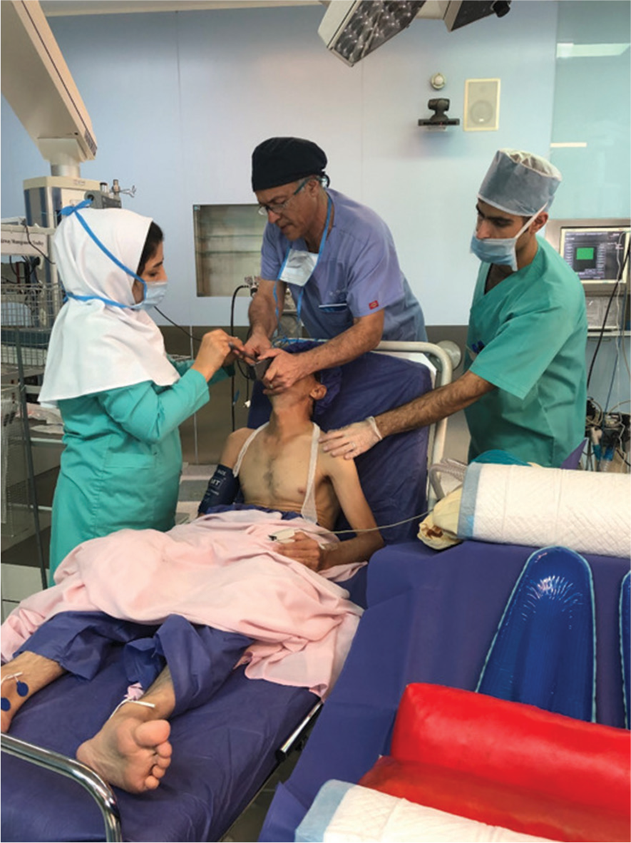
- Induction of anesthesia in our patient with severe fixed thoracolumbar kyphosis, which was carried out in this unusual sitting position.
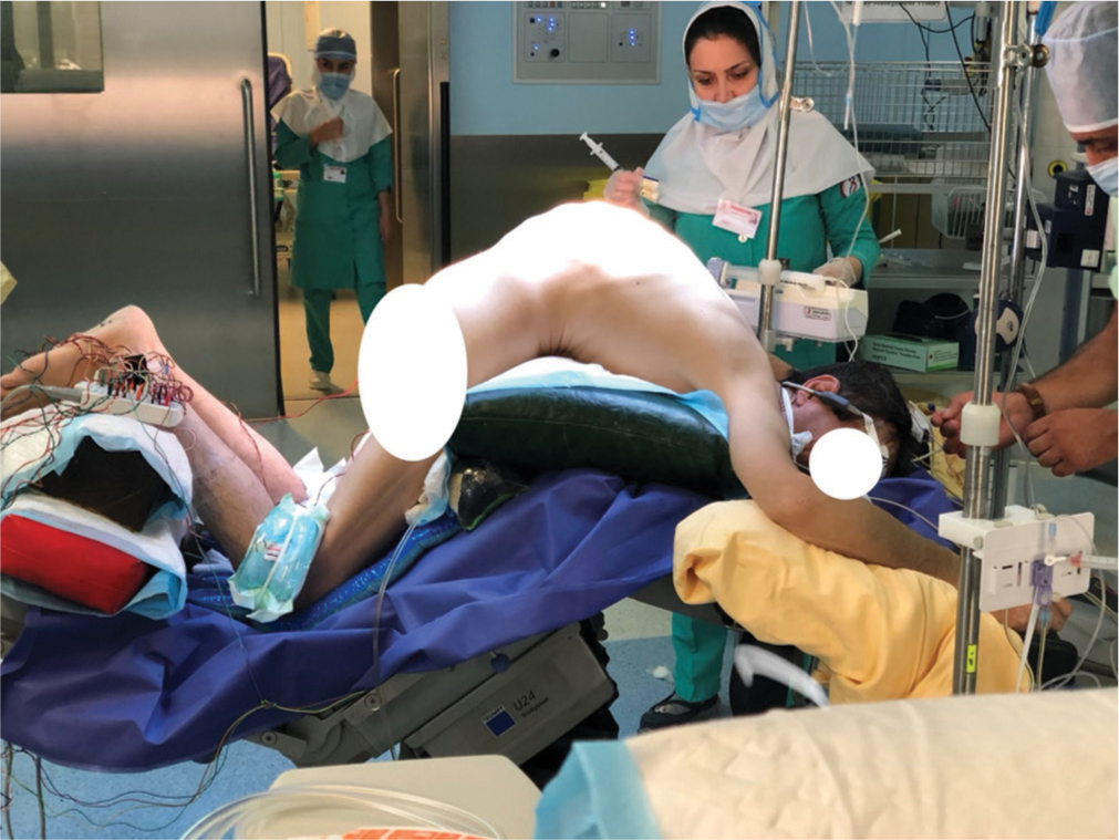
- Proper positioning of the patient with severe fixed thoracolumbar kyphosis required well padding of the pressure points, hanging of the abdomen, and protection of the eyes while avoiding inappropriate head and neck positions.
OPERATIVE TECHNIQUE
Before trying to do the osteotomies, pedicular screws should be inserted first, and their proper position should be checked with biplane fluoroscopy. Osteotomies can be carried out at L1 and L3 or T12 and L2, but this is not an absolute rule. Even sacral PSO has been reported to have satisfactory outcomes.[10]
According to mathematical rules, a fixed amount of wedge resected from the base of a construct in opposition to the same amount of wedge resected from the top could displace the sagittal balance of the construct more significantly; therefore, the lower the osteotomy level, the greater correction of the sagittal vertical axis would be achieved. It is obvious that the amount of kyphosis correction is not related to the location of the osteotomy.[11] Bone resection is carried out using rongeur instruments with special caution. In removing the vertebral body, first, bluntly dissect and displace segmental vessels from the bone and then remove the bone bit by bit. Injury to these vessels can lead to uncontrollable bleeding. Surprisingly, relative total intraoperative blood loss in carefully performed PSO could be trivial.[12]
During PSO, special attention should be paid to preserving the anterior vertebral body hinge. If this critical point is damaged, sudden spinal translation may happen and lead to irreversible cord injury. To prevent this catastrophic event, we used a temporary rod on one side of the spine throughout the surgery. We usually change the side of the rod intermittently to be able to proceed with the bone resection without hindrance.
In critical surgery time, blood pressure should be raised to the appropriate level when intending to correct the deformity. During this critical point, severe hypotension, especially if associated with hypothermia, can raise the possibility of neurologic deficit.[13]
POST-OPERATIVE MANAGEMENT AND FOLLOW-UP
In the early post-operative period, intensive patient care is usually required. Beware of delayed drop of hematocrit. Do not start the regular oral diet early and be careful of abdominal cramps or indigestion during this post-operative period. The patient presented here was followed up for eight years [Figure 3]. He was still satisfied with the results, although he complained of occasional back pain and stiffness with a lumbar pain visual analog scale of 2 of 10 and an Oswestry disability index of 28%.[14-16]
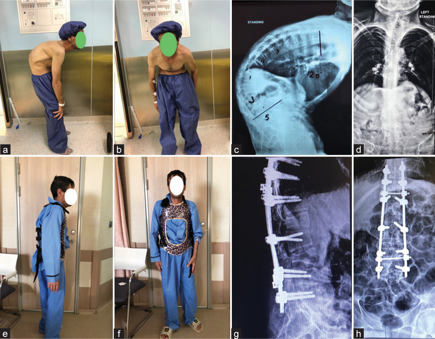
- Pre-operative (a-d) and post-operative (e-h) images of a patient with severe fixed thoracolumbar kyphosis who underwent two-level pedicle subtraction osteotomy (PSO) at L1 and L3. Two-level PSO could efficiently restore center of mass to cone of economy.
DISCUSSION
One-level PSO is usually performed at the L3 level and can correct about 35° of local kyphosis at the lumbar spine segment. However, this type of surgery cannot be effective in severe forms of thoracolumbar kyphosis, and these patients typically need vertebral column resection (VCR) or two-level PSO.[2,3] VCR can be carried out by single-stage posterior or two-stage anterior and posterior approaches. However, it is better reserved for the most severe deformities, especially when they are associated with angular versus round kyphosis. Two-level PSO in severe round thoracolumbar kyphosis not only doubles the strength of a routine PSO correction (to 60° or more) but also harmoniously spreads deformity correction over two separate segments. This great harmonious correction not only decreases the potential risk of neurologic deficit and implant failure but also improves the clinical appearance and energy consumption more efficiently.[3]
Zhang et al. reported surgical outcomes of T12 and L3 (two-level) PSO in nine patients with AS and followed them up for more than 24 months.[2] They reported good clinical appearance in all the cases, with a mean kyphosis correction of 54°. The sagittal vertical axis in their patients improved from 27.3 to 3.4 cm at the last follow-up visit. Similarly, Liu and co-authors, in 2017, in a retrospective clinical study, reported their surgical outcome of two-level PSO in 33 cases with severe kyphosis due to AS.[6] The upper osteotomy site in 10 cases was T12, and in 23 cases, L1. They obtained a kyphosis correction of 26.2° and 27.9° at T12 and L1 osteotomy, respectively, and finally reported satisfactory clinical and radiological outcomes with no significant neurovascular disasters.
CONCLUSION
It is worth noting that these complex surgeries require a teamwork approach between surgeons and anesthesia. This approach can be associated with amazing, satisfactory results if it is done with care for the patient who really needs it and is carefully selected.
ACKNOWLEDGMENT
We are especially grateful to the operating room staff in the spine surgery subspecialty. It would be difficult to perform such precise surgeries without their compassionate cooperation and coordination.
AUTHORS’ CONTRIBUTIONS
FOK contributed to the conception and study design. FOK and GS contributed to data collection, article drafting, and proofreading. Both authors have critically reviewed and approved the final draft and are responsible for the manuscript’s content, accuracy, and similarity index.
ETHICAL APPROVAL
The Institutional Review Board approval is not required.
DECLARATION OF PATIENT CONSENT
The authors certify that they have obtained all appropriate patient consent forms. In the form, the patient has given his consent for his images and other clinical information to be reported in the journal. The patient understands that his name and initials will not be published, and due efforts will be made to conceal his identity, but anonymity cannot be guaranteed.
USE OF ARTIFICIAL INTELLIGENCE (AI)-ASSISTED TECHNOLOGY FOR MANUSCRIPT PREPARATION
The authors confirm that there was no use of artificial intelligence (AI)-assisted technology for assisting in the writing or editing of the manuscript and no images were manipulated using AI.
CONFLICTS OF INTEREST
There are no conflicting relationships or activities.
FINANCIAL SUPPORT AND SPONSORSHIP
This study did not receive any specific grant from funding agencies in the public, commercial, or not-for-profit sectors.
References
- Sagittal spinal alignment in adult spinal deformity: An overview of current concepts and a critical analysis review. JBJS Rev. 2018;6:e2.
- [CrossRef] [PubMed] [Google Scholar]
- Two-level pedicle subtraction osteotomy for severe thoracolumbar kyphotic deformity in ankylosing spondylitis. Eur Spine J. 2014;23:234-41.
- [CrossRef] [PubMed] [Google Scholar]
- Two-level osteotomy for the corrective surgery of severe kyphosis from ankylosing spondylitis: A retrospective series. Spine. 2019;44:1638-46.
- [CrossRef] [PubMed] [Google Scholar]
- Pedicle subtraction osteotomy for the treatment of fixed sagittal imbalance. Surgical technique. J Bone Joint Surg Am. 2004;86-A(Suppl 1):44-50.
- [CrossRef] [PubMed] [Google Scholar]
- Pedicle subtraction osteotomy. JBJS Essent Surg Tech. 2020;10:e0028(1-11).
- [CrossRef] [PubMed] [Google Scholar]
- The safe correction angle of osteotomy at T12 and L1 for ankylosing spondylitis kyphosis: Patients with 2-level osteotomy. Clin Spine Surg. 2017;30:E942-7.
- [CrossRef] [PubMed] [Google Scholar]
- Superior mesenteric artery syndrome after kyphosis correction-A case report. J Orthop Case Rep. 2017;7:67-70.
- [Google Scholar]
- Celiac artery syndrome after correction of kyphoscoliosis. Spine Deform. 2019;7:176-9.
- [CrossRef] [PubMed] [Google Scholar]
- PSO without neuromonitoring: Analysis of peri-op complication rate after lumbar pedicle subtraction osteotomy in adults. Eur Spine J. 2016;25:2629-32.
- [CrossRef] [PubMed] [Google Scholar]
- Sacral pedicle subtraction osteotomy for an extreme case of positive sagittal balance: Case report. J Neurosurg Spine. 2018;28:532-5.
- [CrossRef] [PubMed] [Google Scholar]
- Geometric analysis of pedicle subtraction osteotomy (PSO) for Kyphosis correction: Anterior lengthening may occur at the osteotomized body as well as at the discs above and below. Eur Spine J. 2022;31:2415-22.
- [CrossRef] [PubMed] [Google Scholar]
- Surgical and radiographic outcomes after pedicle subtraction osteotomy according to surgeon's experience. Spine. 2017;42:E795-801.
- [CrossRef] [PubMed] [Google Scholar]
- Raising mean arterial pressure alone restores 20% of intraoperative neuromonitoring losses. Spine. 2018;43:890-4.
- [CrossRef] [PubMed] [Google Scholar]
- A critical review of visual analogue scales in the measurement of clinical phenomena. Res Nurs Health. 1990;13:227-36.
- [CrossRef] [PubMed] [Google Scholar]
- The Oswestry disability index, the Roland-Morris disability questionnaire, and the quebec back pain disability scale: Translation and validation studies of the Iranian versions. Spine. 2006;31:E454-9.
- [CrossRef] [PubMed] [Google Scholar]


