Translate this page into:
A speck of a bone
Corresponding Author:
Ganesh S Dharmshaktu
Department of Orthopaedics, Government Medical College, Haldwani - 263 139, Uttarakhand
India
drganeshortho@gmail.com
| How to cite this article: Dharmshaktu GS. A speck of a bone. J Musculoskelet Surg Res 2021;5:77-78 |
History
A 32-year-old male with a history of a blunt injury to his left foot was advised to have a radiographic examination of the foot, which was reported as no underlying bony injury.
What are your findings?
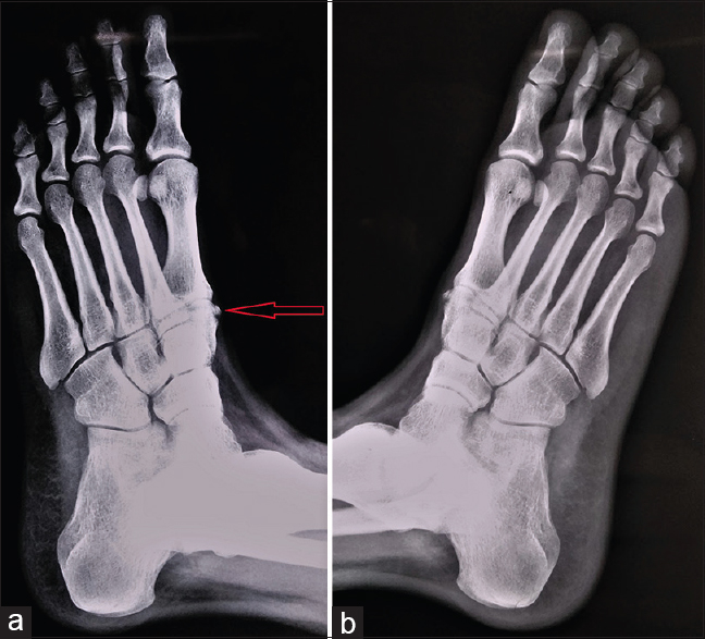
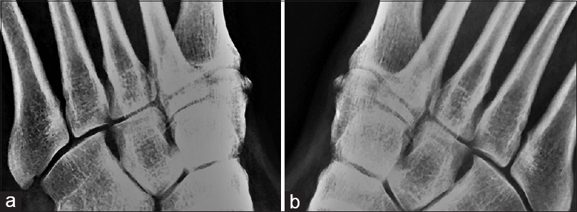
On careful examination, one particular speck of an ossicle was noted medial to the base of the great toe corresponding to the metatarsocuneiform joint [Figure - 1]. The radiograph of the opposite foot confirmed the bilateral presence of the ossicle at an identical site [Figure - 2] on a magnified view.
 |
| Figure 1: An oblique radiographic view of the left (a) and right (b) foot showing an ossicle at identical sites on the medial aspect of the first cuneometatarsal joint |
 |
| Figure 2: The magnified portion of the image showing bilateral presence of the rare “os cuneometatarsale I tibiale” in the left (a) and right (b) foot |
What is your diagnosis?
Os cuneometatarsale I tibiale
Pearls and Discussion
The rare accessory bone at the bilateral medial aspect of the first metatarso-cuneiform joint, mostly an incidental finding, is termed “os cuneometatarsale I tibiale.”[1] Os cuneo-I metatarsale-I plantare is another accessory bone that may mimic cuneometatrsale I tibiale but can be easily differentiated by its location at the plantar surface of the first metatarsocuneiform joint. There are many accessory bones described within the human foot and some of these are more common and well recognized, like accessory navicular, os trigonum, or os vesalianum.[2] Os cuneometatarsale I tibiale has not been routinely described in prevalence studies regarding human accessory bones.[2] The clinical significance of these ossicles is not known due to extreme rarity of their existence. However, these ossicles might occasionally be misdiagnosed with fractures or avulsion injuries by novice practitioners. Acknowledgement and recognition of rare anomalous ossicles are important for proper documentation and further studies.
Declaration of patient consent
The author certifies that he has obtained all appropriate patient consent forms. In the form, the patient has given his consent for his images and other clinical information to be reported in the journal. The patient understands that his name and initials will not be published, and due efforts will be made to conceal his identity, but anonymity cannot be guaranteed.
Financial support and sponsorship
This study did not receive any specific grant from funding agencies in the public, commercial, or not-for-profit sectors.
Conflicts of interest
There are no conflicts of interest.
Further Reading
- Coughlin MJ. Surgery of the foot and ankle in sesamoid and accessory bones of the foot. In: Mann's surgery of the foot and ankle. Vol. 2. Philadelphia: Sanders/Elsevier; 2006. p. 430-94.
- Coskun N, Yuksel M, Cevener M, Arican RY, Ozdemir H, Bircan O, et al. Incidence of accessory ossicles and sesamoid bones in the feet: a radiographic study of the Turkish subjects. Surg Radiol Anat. 2009;31:19-24.
| 1. | Keles-Celik N, Kose O, Sekerci R, Aytac G, Turan A, Güler F. Accessory Ossicles of the Foot and Ankle: Disorders and a Review of the Literature. Cureus. 2017;9:e1881. [Google Scholar] |
| 2. | Rowe SM, Lee KB, Park YB, Bae BH, Kang KD Sesamoids and Accessory Bones of the Forefoot in Normal Korean Adults. J Korean Foot Ankle Soc. 2005;9:20-5. [Google Scholar] |
Fulltext Views
2,099
PDF downloads
1,781


