Translate this page into:
Adequate resection of gigantic cutaneous foot horn associated with a verruca vulgaris
2 Department of Orthopedic Surgery, King Salman Armed Forces Hospital, Tabuk, Saudi Arabia
3 Department of Orthopedic Surgery, King Faisal Specialist Hospital and Research Centre, Riyadh, Saudi Arabia
Corresponding Author:
Sarmad Rashed K. Sulaiman
Department of Orthopaedic Surgery, Al Madina Al Munawara Hospital, 60, Mahzur, Almadinah Almunawwarah 42319
Saudi Arabia
sar.ortsursrm@gmail.com
| How to cite this article: Sulaiman SR, Daghriri Y, Shaheen M, Pant R. Adequate resection of gigantic cutaneous foot horn associated with a verruca vulgaris. J Musculoskelet Surg Res 2021;5:132-135 |
Abstract
Cutaneous horns, also called cornu cutaneum, are a rare disfiguring skin disorder of the keratin, which generally constitutes the skin's superficial layer. They predominantly arise in sunlight-exposed regions of the body and are more common in the elderly. Both males and females are equally affected. They have been named “horns” due to their resemblance to an animals' horns. Cutaneous horns protrude from the skin and can be diagnosed clinically. This case report is about an -84--year-old female who suffered from sole horns, which was treated surgically by marginal excision. The sole's horns had been present for some considerable time, interfered with the patient's walking ability, and made it to a newspaper article even before the patient finally was referred to us. This case report aims to present the surgical management of extraordinarily large-sized foot horn, fortunately, caused by a benign underlying condition, despite typically carries more chances of transformation into malignancy. Up to our knowledge, it would be the largest foot horn documented in the literature.
Introduction
Cutaneous horns are continuing to catch medical practitioners and laypersons' attention with its bizarre and grotesque appearance. The earliest documentation of a cutaneous horn was in 1588 in London, an elderly British woman who appeared in commercial advertisings (as a deviation from the standard).[1]
Everard Home was a surgeon from London, and his notes in 1791 on human cutaneous horns are the earliest mention regarding this anomaly.[1] Farris, an Italian doctor in 1953, was the first who characterized and documented the giant human horns supported with sufficient histology in a case report.[2]
Cutaneous horns are rare, solidly keratinized, finger-like protrusions originating from the skin surface and similar to animal horns. The animal horns are built-up by a well-developed bone at the center covered by the dermal layer and outer hyperkeratotic epidermal superficial layers. There is no bone observed in the gigantic horns in humans.[3] The explanation behind the hyperkeratosis is still undetermined.
A wide range of underlying cutaneous diseases could be associated with cutaneous horns. Particular consideration should, therefore, needs to be paid to any underlying cutaneous disorder and not just to the horn.[4],[5] The risk elements for associated primary cutaneous malignancy include the elderly, male gender, horns with a sizable base (base-to-height proportion), and parts exposed to sunlight.[5],[6] Where there is no associated malignant component, a marginal excision is the mainstay of care. Histopathological examination of the excised lesion is mandatory to rule out any associated pathology.[7]
The aim of this case report to present the surgical management of exceptionally giant foot horn, caused by benign underlying pathology, despite typically carries more chances of transformation into malignancy. Up to our knowledge, it would be the largest foot horn documented in the literature.
Case Report
An 84-year-old female presented to our orthopedic clinic after many years of suffering from large hard protrusions over her right sole. She claimed that it might have started after she stepped over a small piece of metal; months later, she noticed a small projection over a previous localized area of hard sole's skin. The growth of the protrusions was gradual in onset over the years and slowly progressed. With increasing pain and discomfort due to location and size, she could not sleep, and walking was made well nearly impossible. There was no history of discharge from the lesion or related lesions in the different parts of her body. She did not give any history of weight loss and loss of appetite. Her medical history was only significant for hypertension, which was well controlled. She did not give a history of similar lesions in any of her family members.
On examination, the skin was found dry and crusted; there was a solitary, bony hard thickened keratinous mass with five projections arise from the surface of the sole [Figure - 1]. It was firmly adherent to the dermis, and its base measured 8.5 cm in length and 3 cm in width, and the height of the longest projection was 5.5 cm [Figure - 2]. The lesion at its base and the projections were not tender. There was no bleeding, weeping, or discharge from the lesion. Clinically, the skin surrounding the base of the lesion was normal in texture with no infiltrative palpable pathology. There was no associated lymphadenopathy. The foot neurovascular examinations were intact. The remainder of the patient's physical examination was unremarkable. The patient's laboratory tests were within the normal range. Radiography of the foot confirmed that the lesion was arising from the skin of the sole with no deep extension [Figure - 3]. We did not feel that the magnetic resonance image (MRI) study was warranted, based on its very superficial location.
![[Figure - 1]](#fig_SaudiOrthopJ_2021_5_2_132_304228_f1.jpg)
![[Figure - 2]](#fig_SaudiOrthopJ_2021_5_2_132_304228_f2.jpg)
![[Figure - 3]](#fig_SaudiOrthopJ_2021_5_2_132_304228_f3.jpg)
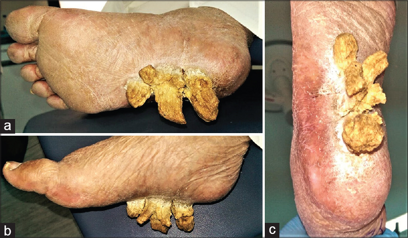 |
| Figure 1 (a-c): A solitary, bony hard thickened keratinous mass with five projections that arise from the surface of the sole. The lesion was of hard consistency and firmly attached to the skin |
 |
| Figure 2 (a-c): The dimensions of the specimen after the excision |
 |
| Figure 3: Radiography of the foot confirmed the lesion was superficial and arising from the skin of the sole with no bony involvement |
For such a large lesion, several punch biopsies of selected areas are appropriate for achieving a diagnosis. After consultation from a dermatologist, we decided to proceed with the excisional biopsy.
With the patient under general anesthesia and a pneumatic tourniquet applied to her thigh, a marginal surgical excision was performed with 5 mm margins [Figure - 4], and the specimen was sent to the histopathology laboratory [Figure - 2]. The base of the lesion was cauterized to reduce the recurrence rate [Figure - 5]. A skin graft was not applied. We preferred the healing by secondary intention with this superficial excision in the sole. The histopathology result was consistent with a verruca vulgaris with clear surgical margins, and no evidence of malignancy was noted. Unfortunately, the patient missed the follow-up after discharge from the hospital.
![[Figure - 4]](#fig_SaudiOrthopJ_2021_5_2_132_304228_f4.jpg)
![[Figure - 5]](#fig_SaudiOrthopJ_2021_5_2_132_304228_f5.jpg)
 |
| Figure 4: The picture showing the marginal excision of the lesion |
 |
| Figure 5 (a,b): The cauterization of the base of the lesion to reduce the recurrence rate |
It should be mentioned here that this case appeared on website news as a social story (dailymail.co.uk, August 7, 2015). The website is a public and not a medical professional website; hence, we did not consider this publication as a case report.[8]
Discussion
Cutaneous horns frequently arise in lightly pigmented skin in people 50 years or over, and they have no sex preference.[9],[10],[11] Although the fact that the pathomechanism for the formation of human horns was not clarified entirely, it was proposed to be because of keratin atypical accumulation and compaction, in addition to its extreme adhesiveness[1] as a response to different underlying cutaneous pathologies that could be benign in 61% of the cases, premalignant in 23.2%, or malignant in 15.7%.[5]
Among the benign primary pathologies are verruca or common warts (as in our patient) commonly caused by human papillomavirus. Viral warts typically exhibit a benign overgrowth of the skin's stratum spinosum layer, numerous papillomas, and hyperkeratosis of the epidermal layer in association with prominent of the papillary capillaries.[12],[13],[14] However, in this benign condition, the triggering factors behind the formation of horns are not identified.[15] Horns that arise from a malignant pathology cannot be differentiated clinically from those having a benign underlying pathology.[16] Tenderness and bleeding at the horn's base are reported to be the signs of malignancy.[16] The presence of inflammation and infiltration surrounding the horn base could be an indication of malignancy.[16]
Where there is a reason to suspect a more aggressive underlying pathology, local imaging should include an MRI and systemic imaging should be mandatory (preferably a positron emission tomography study if available) and the local treatment would entail a wide local excision. A final definitive dermatopathological diagnosis must be obtained.[17]
We searched the PubMed and the Google Scholar websites for similar foot's cutaneous horns, and we found four case report articles;[18],[19],[20],[21] the data were summarized in [Table - 1]. The size of the sole's horn, which is presented in this case report, is the largest.
![[Table - 1]](#tbl_SaudiOrthopJ_2021_5_2_132_304228_t6.jpg)

Superficial biopsies may not reveal a definite diagnosis. Consequently, deep biopsy or complete excision of lesions is advised. Surgical excision remains the treatment of choice in most cases. Marginal excisional biopsy with 3 mm to 1 cm margins is required to deal with the cutaneous horns if associated malignancy is suspected.[1],[16] Other treatment alternatives include cryotherapy (liquid nitrogen), electrocautery, and laser. The cryosurgery freezes and destroys the specimen for histopathology; hence, it is not recommended. These alternative procedures are mainly favored when the lesion is small, with a low grade of suspicion of malignancy. Electrocautery has been applied as an adjuvant to the resection site to minimize the recurrence rates.[1],[9],[16] There was no documentation in the literature about using the chemical cautery, such as phenol solution as an adjuvant to the horn's excision procedure. The recurrence rates are not appropriately documented in the literature, but these lesions respond well to marginal excision.[22],[23]
Declaration of patient consent
The authors certify that they have obtained all appropriate patient consent forms. In the form, the patient has given his consent for his images and other clinical information to be reported in the journal. The patient understands that his name and initials will not be published, and outstanding efforts will be made to conceal his identity, but anonymity cannot be guaranteed.
Financial support and sponsorship
This research did not receive any specific grant from funding agencies in the public, commercial, or not-for-profit sectors.
Conflicts of interest
There are no conflicts of interest.
Authors' contributions
SS conceived and designed the study and wrote the initial draft of the article. SS and YD collected, organized, analyzed, and interpreted the data. MS and RP provided critical revision and wrote the final draft of the article. All authors have critically reviewed and approved the final draft and are responsible for the manuscript's content and similarity index.
| 1. | Shahi S, Bhandari TR, Pantha T. Verrucous carcinoma in a giant cutaneous horn: A case report and literature review. Case Rep Otolaryngol 2020;2020:7134789. [Google Scholar] |
| 2. | Farris G. Histological considerations on a case of a voluminous cutaneous horn. Minerva Dermatol 1953;28:159-65. [Google Scholar] |
| 3. | Michal M, Bisceglia M, Di Mattia A, Requena L, Fanburg-Smith JC, Mukensnabl P, et al. Gigantic cutaneous horns of the scalp: Lesions with a gross similarity to the horns of animals: A report of four cases. Am J Surg Pathol 2002;26:789-94. [Google Scholar] |
| 4. | Akan M, Yildirim S, Avci G, Aköz T. Xeroderma pigmentosum with a giant cutaneous horn. Ann Plast Surg 2001;46:665-6. [Google Scholar] |
| 5. | Yu R, Pryce DW, Macfarlane AW, Stewart TW. A histopathological study of 643 cutaneous horns. Br J Dermatol 1991;124:449-52. [Google Scholar] |
| 6. | Fox GN. Facial lesion that came “out of nowhere.” J Fam Pract. 2004;53:779-81. [Google Scholar] |
| 7. | Solanki LS, Dhingra M, Raghubanshi G, Thami GP. An innocent giant. Indian J Dermatol 2014;59:633. [Google Scholar] |
| 8. | Available from: https://www.dailymail.co.uk/health/article-3189480/Agony-pensioner-three-TOENAIL-HORNS-growing-foot.html. [Last accessed on 2020 Jul 10]. [Google Scholar] |
| 9. | Fernandes NF, Sinha S, Lambert WC, Schwartz RA. Cutaneous horn: A potentially malignant entity. Acta Dermatovenerol Alp Pannonica Adriat 2009;18:189-93. [Google Scholar] |
| 10. | Copcu E, Sivrioglu N, Culhaci N. Cutaneous horns: Are these lesions as innocent as they seem to be? World J Surg Oncol 2004;2:18. [Google Scholar] |
| 11. | Oludiran OO, Ekanem VJ. Cutaneous horns in an African population. J Cutan Aesthet Surg 2011;4:197-200. [Google Scholar] |
| 12. | Dyall-Smith D, Mason G. Plantar wart presenting as a cutaneous horn. Australas J Dermatol 1995;36:214-5. [Google Scholar] |
| 13. | Wang W, Wang C, Xu S, Chen C, Tong X, Liang Y, et al. Detection of HPV-2 and identification of novel mutations by whole genome sequencing from biopsies of two patients with multiple cutaneous horns. J Clin Virol 2007;39:34-42. [Google Scholar] |
| 14. | Croissant O, Breitburd F, Orth G. Specificity of cytopathic effect of cutaneous human papillomaviruses. Clin Dermatol 1985;3:43-55. [Google Scholar] |
| 15. | Solanki LS, Dhingra M, Raghubanshi G, Thami GP. An innocent giant. Indian J Dermatol 2014;59:633. [Google Scholar] |
| 16. | Tambe K, Reuser TT, Sampath RG, Burns J, Ahluwalia H, Bowyer JD, et al. A prospective, multicentre study of malignant and premalignant lesions at the base of periocular cutaneous horns. Orbit 2012;31:404-7. [Google Scholar] |
| 17. | Sathyanarayana SA, Deutsch GB, Edelman M, Cohen-Kashi KJ. Cutaneous horn: A malignant lesion? A brief review of the literature. Dermatol Surg 2012;38:285. [Google Scholar] |
| 18. | Singh O, Nair V, Ranjan P, Gaba S, Singh K. Cutaneous Horns: enigma–Remembering Lady Dimanche. Sri Lanka Journal of Surgery. 2019;37. [Google Scholar] |
| 19. | Ingram NP. Cutaneous horns: A review and case history. Ann R Coll Surg Engl 1978;60:128-9. [Google Scholar] |
| 20. | Lalehparvar S, Mohiuddin A, Labib I. Successful excision of cutaneous horns in the foot and application of a full-thickness pinch skin graft for primary closure. J Am Podiatr Med Assoc 2017;107:158-61. [Google Scholar] |
| 21. | Dyall-Smith D, Mason G. Plantar wart presenting as a cutaneous horn. Australas J Dermatol 1995;36:214-5. [Google Scholar] |
| 22. | Mencía-Gutiérrez E, Gutiérrez-Díaz E, Redondo-Marcos I, Ricoy JR, García-Torre JP. Cutaneous horns of the eyelid: A clinicopathological study of 48 cases. J Cutan Pathol 2004;31:539-43. [Google Scholar] |
| 23. | Pyne J, Sapkota D, Wong JC. Cutaneous horns: Clues to invasive squamous cell carcinoma being present in the horn base. Dermatol Pract Concept 2013;3:3-7. [Google Scholar] |
Fulltext Views
2,456
PDF downloads
542


