Translate this page into:
Case of ankle pain
Corresponding Author:
Nizar A Al-Nakshabandi
Department of Radiology, College of Medicine, King Saud University, Riyadh
Saudi Arabia
nizar97@hotmail.com
| How to cite this article: Al-Nakshabandi NA. Case of ankle pain. J Musculoskelet Surg Res 2020;4:168-169 |
History
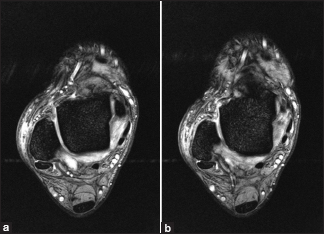
A 27-year-old female presented with pain and swelling. She gave a history of twisting ankle injury (inversion with plantar flexion). The swelling is more on the anterolateral aspect of the ankle. Her radiographs were unremarkable. She had a magnetic resonance imaging (MRI) that is shown here:
- What are your findings?
- What is the differential diagnosis?
- What are the causes?
Findings
Axial T2-weighted images at the level of the anterior talofibular ligament (ATFL) demonstrate an absent ATFL; instead, only fluid is seen at the site of its attachment. Also, a torn anterior inferior tibiofibular ligament is shown.
Differential Diagnosis
- ATFL tear
- Anterolateral impingement syndrome of the ankle.
Diagnosis
- Complete tear of the ATFL associated with anterior inferior tibiofibular ligament tear.
Pearls and Discussion
Ankle ligaments
Lateral complex
- ATFL
- Posterior talofibular ligament
- Calcaneofibular ligament.
Medial complex (deltoid)
- Tibionavicular ligament
- Tibiospring ligament
- Tibiocalcaneal ligament
- Anterior tibiotalar ligament
- Posterior tibiotalar ligament.
Ankle syndesmosis
- Anterior inferior tibiofibular ligament
- Posterior inferior tibiofibular ligament
- Inferior transverse ligament
- Distal interosseous ligament or membrane.
Spring ligament complex (calcaneonavicular ligament)
- Superomedial calcaneonavicular ligament
- Medioplantar oblique calcaneonavicular ligament
- Inferoplantar longitudinal calcaneonavicular ligament.
Lateral ligament complex is the most commonly injured group of ankle ligaments and is often associated with other ligamentous injuries in the ankle. The lateral complex compromising the ATFL, the calcaneofibular ligament, and the posterior talofibular ligament is adequately managed with routine axial MRI.
Grading
The severity of the ligamentous injury can be classified into three grades:
- Grade I: Injury without macroscopic tears
- Grade II
- Partial tear
- Mild-to-moderate joint instability may be present.
- Grade III
- Complete tear
- Inability to weight-bear
- Associated with significant joint instability.
Mechanism of injury is usually inversion with plantar flexion to the ankle.
Two-thirds of the injuries are isolated ATFL.
Declaration of patient consent
The author certifies that he has obtained all appropriate patient consent forms. In the form, the patient has given her consent for her images and other clinical information to be reported in the journal. The patient understands that her name and initials will not be published and due efforts will be made to conceal her identity, but anonymity cannot be guaranteed.
Financial support and sponsorship
Nil.
Conflicts of interest
There are no conflicts of interest.
Further Reading
- Haraguchi N, Toga H, Shiba N, Kato F. Avulsion fracture of the lateral ankle ligament complex in severe inversion injury: Incidence and clinical outcome. Am J Sports Med 2007;35:1144-52.2.
- Kumai T, Takakura Y, Rufai A, Milz S, Benjamin M. The functional anatomy of the human anterior talofibular ligament in relation to ankle sprains. J Anat 2002;200:457-65.
- Lee MH, Cha JG, Lee YK, Choi GC, Paik SH, Lee HK, et al. The bright rim sign on MRI for anterior talofibular ligament injury with arthroscopic correlation. AJR Am J Roentgenol 2012;198:885-90.
Fulltext Views
1,391
PDF downloads
534


