Translate this page into:
Case of bizarre painless finger swelling
Corresponding Author:
Leon Alexander
Department of Surgery, Division of Plastic Surgery, Sheikh Khalifa Medical City, Abu Dhabi
UAE
dr.leonalex@gmail.com
| How to cite this article: Alexander L. Case of bizarre painless finger swelling. J Musculoskelet Surg Res 2021;5:136-137 |
History
A 25-year-old-male patient, right-hand dominant, a welder by profession, presented with a 2-year history of swelling of the right middle finger. There is a slow, gradual increase in the size of the swelling over time with no previous history of trauma. He has no associated symptoms of neurovascular compression or reduced range of motion.
- What are your findings?
- What is the differential diagnosis?
- What is the treatment?
Findings
The anteroposterior view [Figure - 1]b and oblique view [Figure - 1]a of the right hand show a well-defined, wide-based bony growth arising from the metaphysis of the proximal phalanx of the right middle finger. This mineralizing exophytic lesion arises from the cortex of the proximal phalanx with no medullary involvement. The flecks of calcification within the tumor are pretty characteristic. It may or may not be associated with underlying cortical irregularity. There is no associated osteolysis, cortical flaring, or periosteal reaction.
![[Figure - 1]](#fig_SaudiOrthopJ_2021_5_2_136_315679_f1.jpg)
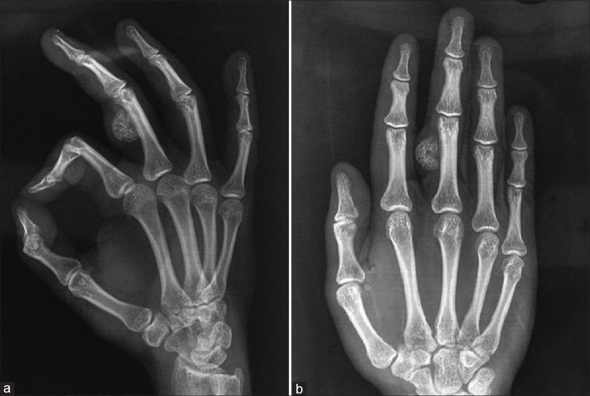 |
| Figure 1: X-ray of the right hand anteroposterior view (b) and oblique view (a) showing an exophytic osteochondromatous lesion arising from the proximal phalanx of the middle finger |
Differential Diagnosis
- Osteochondroma
- Periosteal chondroma
- Parosteal osteosarcoma
- Periosteal osteosarcoma
- Periosteal chondrosarcoma
- Florid reactive periostitis
- Myositis ossificans
- Turret exostosis.
Diagnosis
Bizarre parosteal osteochondromatous proliferation (BPOP - Nora lesion).
Pearls and Discussion
Nora lesion or BPOP was first described by an American pathologist, Nora et al. He reported 35 cases of BPOP occurring in the proximal phalanges, metatarsals, or metacarpals of the hands and feet.[1]
BPOP is a rare, benign, exostotic osteochondromatous tumor representing a diagnostic challenge due to its clinical and radiological similarity with osteochondromas.
Radiological findings are nonspecific, but computed tomography or magnetic resonance imaging may help identify this tumor. It can be differentiated from osteogenic tumors by the absence of cortical flaring and chondromatous tumors by the lack of medullary continuity.[2]
Definitive diagnosis is established by histology, which shows a mixture of bone, cartilage, and a fibrous myxoid spindle cell stroma without cellular atypia. A characteristic feature is the distinct blue tinctorial characteristic of the bone in the lesion.[3]
The treatment is surgical excision. However, recurrence is common. They can be locally invasive but do not metastasize. It has been suggested for lesions presenting early to do surgical excision of the tumor, pseudocapsule and periosteal tissue, and decortication of bony irregularities to minimize recurrence. For a late presentation with locally aggressive tumors, the treatment of choice is wide local resection and/or ray amputation.[1],[2],[3]
Declaration of patient consent
The author certifies that he has obtained all appropriate patient consent forms. In the form, the patient has given his consent for his images and other clinical information to be reported in the journal. The patient understands that his name and initials will not be published and due efforts will be made to conceal his identity, but anonymity cannot be guaranteed.
Financial support and sponsorship
This research did not receive any specific grant from funding agencies in the public, commercial, or not-for-profit sectors.
Conflicts of interest
There are no conflicts of interest.
| 1. | Nora FE, Dahlin DC, Beabout JW. Bizarre parosteal osteochondromatous proliferations of the hands and feet. Am J Surg Pathol 1983;7:245-50. [Google Scholar] |
| 2. | Gursel E, Jarrahnejad P, Arneja JS, Malamet M, Akinfolarin J, Chang YJ. Nora's lesion: Case report and literature review of a bizarre parosteal osteochondromatous proliferation of a small finger. Can J Plast Surg 2008;16:232-5. [Google Scholar] |
| 3. | Meneses MF, Unni KK, Swee RG. Bizarre parosteal osteochondromatous proliferation of bone (Nora's lesion). Am J Surg Pathol 1993;17:691-7. [Google Scholar] |
Fulltext Views
1,423
PDF downloads
459


