Translate this page into:
Complex forearm deformities in brachial plexus birth palsy: Radial head dislocation, forearm bones crossing, and distal ulna volar dislocation
*Corresponding author: J. Terrence Jose Jerome, Department of Orthopaedics, Hand and Reconstructive Microsurgery, Olympia Hospital and Research Centre, Trichy, Tamil Nadu, India. terrencejose@gmail.com
-
Received: ,
Accepted: ,
How to cite this article: Jerome JTJ. Complex forearm deformities in brachial plexus birth palsy: Radial head dislocation, forearm bones crossing, and distal ulna volar dislocation. J Musculoskelet Surg Res. 2024;8:403-5. doi: 10.25259/JMSR_216_2024
Abstract
We present a rare and complex case involving a 6-year-old boy with brachial plexus birth palsy, which is complicated by secondary developmental changes such as proximal radial head dislocation, distal ulna dislocation, and a severe supination deformity in the forearm. Despite the severity of his condition, his parents declined further treatment. This case underscores the presence of unclassifiable complex anomalies and highlights the need to expand the current classification system, especially when these anomalies are associated with birth palsy. Our report emphasizes the importance of recognizing and addressing these intricate presentations to enhance diagnostic and therapeutic strategies.
Keywords
Brachial plexus
Birth palsy
Dislocation
Distal ulna
Radial head
Supination deformity
INTRODUCTION
Brachial plexus birth palsy often leads to supination deformity due to muscle imbalance, particularly affecting the C6–8 region.[1-3] This imbalance results from the impaired functioning of the median nerve, impacting muscles such as pronator teres and quadratus. Concurrently, the biceps muscle, acting as a supinator, can cause contracture, leading to anterior dislocation of the radial head and forearm supination deformity.[1] Muscle imbalances may further shrink the interosseous membrane, exacerbating the supination contracture. However, distal radioulnar joints generally remain unaffected.
We present a distinctive case of brachial plexus birth palsy, showcasing anterior dislocation of the radial head, crossing of forearm bones, volar dislocation of the distal ulna, and a severe supination deformity. This rare manifestation emphasizes the importance of recognizing diverse sequelae in brachial plexus birth palsy for enhanced comprehension of pathophysiology and treatment considerations.
CASE REPORT
A 6-year-old boy, born with brachial plexus birth palsy, sought consultation at our hand clinic due to a prominent deformity in his left upper limb. Initial management involved therapy and splinting, steering clear of surgical interventions. Physical examination revealed a severe supination deformity in the left forearm and a flail hand [Figure 1]. The British Medical Research Council muscle grade indicated strength at 4/5 in the shoulder and elbow. A scapulothoracic deformity was evident, marked by a hypoplastic, elevated, and rotated scapula.

- The clinical picture shows a severe supination deformity in the left forearm.
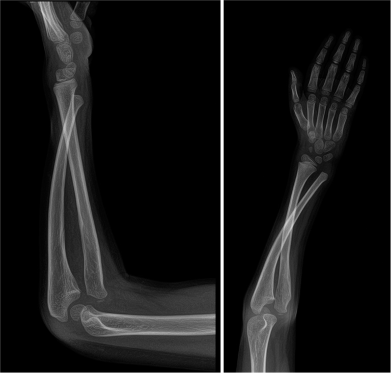
While the shoulder and elbow displayed a full range of motion, rotations at the elbow and forearm/wrist were restricted, resulting in a severe supination deformity. The hand exhibited a flail state with no active finger movements, significantly limiting bimanual activities and daily hand usage. Radiographic studies uncovered anterior dislocation of the radial head, a crossing of the radius and ulna in the forearm, and volar dislocation of the distal ulna [Figure 2]. Despite the severity of the condition, surgical interventions such as corrective osteotomy and biceps re-routing were proposed. However, the parents declined these treatments. The exact reasons for their decision to decline surgery remain unclear.

- The radiographs show anterior dislocation of the radial head and volar dislocation of the distal ulna with a crossing of both forearm bones.
DISCUSSION
This case highlights a rare deformity associated with brachial plexus birth palsy. It emphasizes the importance of considering supination with radial head and distal ulnar dislocation, along with crossing forearm bones. Although challenging to treat, understanding the pathophysiology becomes imperative for surgeons.
Addressing supination contractures, colloquially termed “begging hand,” involves corrective osteotomy and brachioradialis/biceps re-routing, enhancing hand positioning and appearance.[1-3] Severe contractures may necessitate interosseous membrane release and radial osteotomy. However, attempts at open reduction for radial head dislocation have proven less successful.[2]
The rarity of crossing forearm bones poses additional challenges, potentially linked to interosseous membrane shrinkage and biceps contracture, contributing to supination deformity. Imbalanced muscular contractions may impact joint surface development, potentially causing instability as the child grows.[3] Persistent volar dislocation of the distal ulna, attributed to incongruent articular surfaces, could be mitigated through corrective osteotomy and stabilization techniques.
While parents declined surgery in our case, surgeons must inform them about diverse treatment options, potential complications, and future considerations like tendon transfer techniques.
This case highlights the presence of complex, unclassifiable anomalies and underscores the need to expand the current classification system, especially when these anomalies are associated with birth palsy. Our report aimed to emphasize the importance of recognizing and addressing these intricate presentations to enhance diagnostic and therapeutic strategies.
CONCLUSION
This case contributes a rare variant involving distal ulna volar dislocation and crossing forearm bones to the spectrum of the brachial plexus birth palsy sequelae. Every hand surgeon should be cognizant of such variations. In our instance, parental reluctance for surgery hindered potential improvements in hand appearance, emphasizing the importance of comprehensive patient education and shared decision-making.
ETHICAL APPROVAL
Institutional Review Board approval is not required.
DECLARATION OF PATIENT CONSENT
The author certifies that he has obtained all appropriate patient consent forms. In the form, the patient’s parents have given their consent for the patient’s images and other clinical information to be reported in the journal. The parents understand that the patient’s name and initials will not be published, and due efforts will be made to conceal his identity, but anonymity cannot be guaranteed.
USE OF ARTIFICIAL INTELLIGENCE (AI)-ASSISTED TECHNOLOGY FOR MANUSCRIPT PREPARATION
The author confirms that there was no use of artificial intelligence (AI)-assisted technology for assisting in the writing or editing of the manuscript and no images were manipulated using AI.
CONFLICTS OF INTEREST
There are no conflicting relationships or activities.
FINANCIAL SUPPORT AND SPONSORSHIP
This study did not receive any specific grant from funding agencies in the public, commercial, or not-for-profit sectors.
References
- Surgical correction of supination deformity in children with obstetric brachial plexus palsy. J Hand Surg Br. 2002;27:20-3.
- [CrossRef] [PubMed] [Google Scholar]
- Radial head dislocation as a rare complication of obstetric brachial plexus palsy: Literature review and five case series. Hand Surg. 2012;17:33-6.
- [CrossRef] [PubMed] [Google Scholar]
- The supination deformity and associated deformities of the upper limb in severe birth lesions of the brachial plexus. J Bone Joint Surg Br. 2009;91:511-6.
- [CrossRef] [PubMed] [Google Scholar]


