
Translate this page into:
Effects of proprioceptive neuromuscular facilitation technique on scapular dyskinesis in patients with subacute stroke
*Corresponding author: Moquddas Gull, Department of Physiotherapy, Bin Zahid Medical Complex, Lahore, Pakistan. moquddasphysio17@gmail.com
-
Received: ,
Accepted: ,
How to cite this article: Abdul Rahman R, Sattar H, Zulfiqar A, Shakil Butt B, Shakir S, Fatima N, et al. Effects of proprioceptive neuromuscular facilitation technique on scapular dyskinesis in patients with subacute stroke. J Musculoskelet Surg Res. 2024;8:125-32. doi: 10.25259/JMSR_267_2023
Abstract
Objectives:
The objective of this was to compare the effects of the proprioceptive neuromuscular facilitation (PNF) hold-relax technique on pain severity, range of motion (ROM) of the shoulder, shoulder disability, and scapular asymmetry in stroke patients with scapular dyskinesis.
Methods:
The study used a randomized clinical trial design including 46 patients ranging from 40 to 60 years with at least five months of stroke with type-1 scapular dyskinesia. The participants were divided into two groups, taking the upper limb Diagonal 1 (D1) flexion pattern and Diagonal 2 (D2) flexion pattern, respectively, which were allocated by consecutive sampling using the lottery method. We used a visual analog scale (VAS) for pain, goniometry for shoulder ROM, shoulder pain and disability index (SPADI) for shoulder disability, and lateral scapular slide test for scapular asymmetry.
Results:
D1 flexion and D2 flexion both improved the ROM in both groups (P < 0.05) and decreased pain and disability, while in-between comparisons did not find a significant difference between the effectiveness of both treatments in terms of pain measured by VAS, ROM, that is, flexion, extension, and abduction (P > 0.05). In terms of external and internal rotation, the D1 flexion pattern of PNF techniques showed more improvement as compared to D2 flexion (P < 0.05). While in terms of SPADI, D2 flexion showed more improvement as compared to D1 flexion (P < 0.05).
Conclusion:
Scapular PNF substantially influences stroke patients’ shoulder discomfort and ROM.
Keywords
Hold-relax
Proprioceptive neuromuscular facilitation
Scapular stabilization exercise
Scapular dyskinesis
Stroke

INTRODUCTION
The scapula is a triangular-shaped flat bone located on the back side of the thorax, which plays a key role in facilitating shoulder movement and maintaining stability.[1,2] One of the primary movements associated with the scapula is upward rotation. This movement is crucial for activities that require lifting the arm overhead such as reaching for objects on high shelves or performing overhead sports movements.[3,4] The physiology of the scapula involves the coordinated actions of various muscles and ligaments that attach to its distinct features including the borders, angles, and prominent processes.
Scapular pathophysiology can result from various factors including injury, poor posture, muscle weakness, and nerve damage. These factors can contribute to structural and functional abnormalities in the scapula leading to altered movement patterns and biomechanics in the shoulder joint. One common condition associated with scapular pathophysiology is scapular dyskinesis. It can also contribute to conditions such as rotator cuff impingement and shoulder bursitis. Another prevalent scapular pathology is scapular winging. Scapular winging is commonly observed in individuals involved in activities that require repetitive pushing or lifting motions.[3]
Physical therapy plays a central role in rehabilitating scapular disorders. Therapeutic interventions aim to restore normal scapular position, movement, and muscle strength. Specific exercises are prescribed to target the scapular stabilizing muscles including the trapezius, serratus anterior, and rhomboids. These exercises help to strengthen weak muscles, improve muscle balance, and promote proper scapular kinematics. Techniques used in manual treatment such as releasing trigger points and mobilizing joint and soft tissue may also be used to address muscle imbalances, restore normal alignment, and reduce pain. Proprioceptive exercises, balance training, and kinesthetic awareness drills are employed to improve muscle activation and coordination enabling individuals to perform functional movements with optimal scapular control.
In some cases, bracing or taping techniques may be used to provide external support and promote correct scapular positioning during activities. These assistive devices can help individuals maintain proper scapular alignment and prevent excessive or abnormal movements.[5]
Stroke is a clinical syndrome characterized by poor blood flow to the brain that leads to the sudden death of brain cells in localized areas and the formation of a long-lasting, focal neurological impairment as a result of a vascular incident. As toxic metabolites accumulate in the body, the brain parenchyma gets damaged structurally. After a stroke, a low-tone flaccid stage without voluntary control followed by the spastic stage, which results in scapulothoracic joint asymmetry, are the initial symptoms.[6,7] The occurrence of hemiplegic shoulder pain is a widely recognized and significant complication that can arise in the aftermath of a stroke. Its prevalence has been reported to range from 34% to 84% among stroke patients making it a common and debilitating issue.[6] Age plays a significant role, as the incidence of strokes tends to rise as individuals get older. The constant high pressure within the blood vessels can weaken and damage them over time increasing the likelihood of a stroke. Smoking and high cholesterol levels also increase the risk of stroke.[8] The occurrence of hemiplegic shoulder pain is a widely recognized and significant complication that can arise in the aftermath of a stroke.
Proprioceptive neuromuscular facilitation (PNF) is a stretching technique that involves using specific patterns of movement and resistance to improve muscle strength, range of motion (ROM), and function. The PNF is often used in the rehabilitation of stroke patients to address muscle weakness and impaired motor control. The PNF techniques involve various movements such as diagonal movement patterns, rhythmic stabilization, and repeated contractions. These movements are performed with resistance provided by the therapist or equipment.[9] The hold-relax approach is a type of PNF frequently used to treat muscular tightness, stiffness, and restricted ROM. Muscle deficits in stroke survivors can be caused by a variety of variables such as muscle spasticity and diminished control over their movements. These disorders can lead to muscular imbalances, reduced flexibility, and difficulties performing functional motions with the afflicted limbs. The technique involves actively contracting a muscle for several seconds followed by a brief period of relaxation and then a passive stretch of the muscle. Stroke rehabilitation seeks to recover motor function and functional capacities following a stroke.[10]
In clinical practice, most stroke patients were treated in all aspects but mostly ignored scapular mechanics. Therefore, an untreated scapular dyskinesis may lead to shoulder pain and dysfunction, so there was a need to design an alternate plan of care if routine management failed, which would focus on scapular mechanics. For its treatment, PNF hold-relax with conservative treatment such as ROM and strength, which were effective, but there was limited literature on combined impact. Consequently, studies investigating these interventions in terms of scapular alignment were required. The present study aimed to see the effects of the PNF hold-relax technique on scapular dyskinesis in patients with subacute stroke a nd to compare the effects of Diagonal 1 (D1) and Diagonal 2 (D2) flexion patterns of PNF on pain, ROM, disability, and scapular asymmetry in patients with subacute stroke. The findings of this study would help the physiotherapist design the plan of care for scapular dyskinesis among patients, who underwent PNF hold-relax technique and scapular stabilization exercise. These results would be utilized by researchers in future studies. Patients would use the findings to receive a cost-effective treatment, as these interventions can be performed individually.
MATERIALS AND METHODS
This study used a randomized clinical trial. It was conducted at Ibn-e-Siena Hospital and Research Institute, Multan, Pakistan. The duration of the study was six months. A convenient sampling technique was used for the selection of the sample. The sample size of the study was 46.
The sample size was calculated using the formula given by Charan and Biswas.[11]
SD: Standard deviation, Z: Standard normal deviate, α: Type I error, β: Type II error, d: Precision.
The sample size was calculated from Joshi et al.[12] by taking the visual analog scale (VAS) as an outcome measure of interest. A total of 46 subjects were recruited into the study after screening through the selection criteria [Table 1].
| Mean VAS of Group A=4.25 |
| Mean VAS of Group B=3.28 |
| SD=0.93 |
| Zα/2 type I error of 5%=1.96 |
| Zβ keeping power of study at 80%=0.84 |
| Keeping allocation ratio 1:1 |
| Sample size=22.87 approximately 23/group |
VAS: Visual analog scale, SD: Standard deviation
The study’s inclusion criteria were stroke patients with an onset of at least five months. Patients aged 40–60 years of age included both males and females. Patients developing unilateral scapular dyskinesis due to stroke were assessed by lateral scapular slide test (LSST) and typed 1 scapular dyskinesia prominent infernomedial border of the scapula. Exclusion criteria of the study included a neurological deficit of the upper extremity, any surgery of the upper extremity, active infections, hypermobility, calcification of the soft tissues, fragile skin, musculoskeletal injuries as upper limb fractures, dislocation, joint instability, or any soft-tissue injuries on the affected side, etc., other illnesses such as supraspinatus, or bicep tendinitis, fibromyalgia, frozen shoulder, and arthritis that produced discomfort in the upper extremities, etc., and severe depression.
The data was collected through a questionnaire, which consisted of socioeconomic data that was recorded before the study. The participants were divided into two groups taking the upper limb D1 flexion pattern as group 1 and the D2 flexion pattern as group 2, allocated by consecutive sampling technique by lottery method. The baseline treatment, moist hot-pack, and scapular stabilization exercise were done for both groups undergoing the same protocol of 12 sessions (three sessions/week). The measurements of the study parameters including pain and shoulder ROM were accomplished at three time periods, that is, at baseline, after second week, and after fourth week. Shoulder disability was assessed by shoulder pain and disability index (SPADI) at baseline and after four weeks.
The patients were given the baseline treatment before respective treatment as per the groups allocated to them including treatment with a moist hot pack and scapular stabilization exercises. The moist hot pack was applied to the affected site of the shoulder. Scapular stabilization consists of scapular retraction, external rotation, shoulder diagonals, horizontal rows, shoulder extension, and prone horizontal extension exercises. Each exercise was repeated with two sets of 10 repetitions. The time for performing baseline treatment was 25 min.
After the baseline treatment, group A patients received the scapular hold-relax technique applied in the upper limb D1 flexion pattern [Figure 1]. The antagonistic pattern in D1 is a flexion-adduction-external rotation that includes two sets of 20 repetitions performed with a 20-s rest period between the sets. The patient was lying on the unaffected side while the therapist stood in line with the desired motion. The first therapist gave preparatory instructions to patients. In this exercise, the therapist’s proximal hand grasps the patient’s upper arm on the anteromedial surface exerting resistance in the opposite direction of the movement. Simultaneously, the distal hand holds the patient’s hand with the fingers positioned on the ulnar side and the thumb on the radial side allowing the patient’s wrist to flex toward the radial side. This particular hand positioning technique is referred to as the lumbrical grip.[13] The total time for baseline treatment and D1 flexion exercise was at least 50 min.
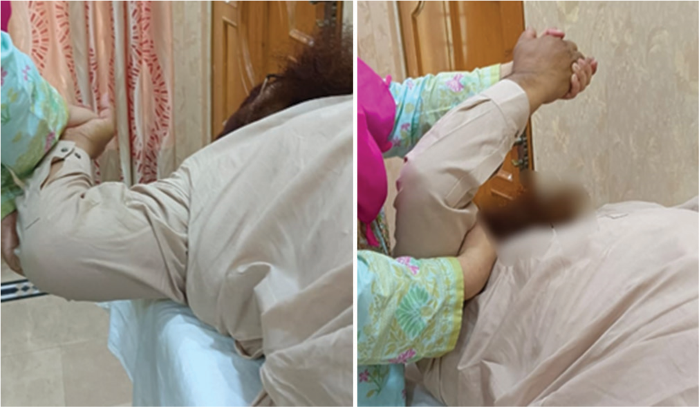
- Diagonal 1 flexion pattern.
After the baseline treatment, the patients of group B received a scapular hold-relax technique applied in the upper limb D2 flexion pattern [Figure 2]. The antagonistic pattern in D2 is flexion abduction-external rotation that included two sets of 20 repetitions performed with 20 s rest period between sets. The patient was lying on the unaffected side as the therapist made the desired motion while standing in line. The first therapist gave preparatory instructions to patients. The patient’s hand’s dorsal-radial surface is held in the therapist’s distal hand. To provide pressure in the opposite direction of movement, the proximal hand grasps the anterior-lateral surface of the upper arm from below.[13] The total time for baseline treatment and D2 flexion exercise was at least 50 min.
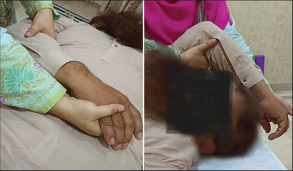
- Diagonal 2 flexion pattern.
The primary outcomes of the study were pain and shoulder asymmetries. The assessment of pain severity and scapular asymmetry was used to measure the shoulder’s ROM (flexion, extension, abduction, internal rotation, and external rotation), the VAS LSST, and Goniometry, respectively. The VAS was used to assess the characteristics of pain that a patient is experiencing. This scale is the gold standard test.[2] The LSST was utilized to assess scapular asymmetries under various load conditions. Three test positions were applied using Kibler’s approach. The LSST had good reliability values. The inferior angle of the scapula and the spinous process are identical in all three locations in this technique when evaluated bilaterally with a piece of tape in the horizontal plane. If it demonstrates a difference of 1.5 cm or more in any of the three places, it was determined that the LSST has a positive result.[14] Shoulder disability was a secondary outcome measure. The SPADI evaluates pain as well as the other upper extremity functional activities. The SPADI has high reliability and validity.[15]
Statistical analysis
The Statistical Package for the Social Sciences version 26 was used for the analysis and interpretation of results. The normality of the data was assessed through Shapiro–Wilk statistical tests. The VAS and shoulder ROM were violating the normal distribution (P < 0.05), so a non-parametric test (Friedman Test and Mann–Whitney U-test) was used for the analysis of VAS and shoulder ROM. The SPADI followed the normal distribution (P > 0.05), so parametric tests (Repeated measure analysis of variance and independent t-test) were used for its analysis.
RESULTS
Descriptive statistics
The demographic statistics of the study is shown in Table 2. The mean age of participants in group A (D1 flexion) was 48.65 ± 5.21, while in group B (D2 flexion), it was 28.21 ± 6.32. The age range for group A (D1 flexion) is between 42 and 58 years old. The age range for group B (D2 flexion) is between 40 and 57 years old. Among 46 participants in the research, 24 were female (52.2%), while 22 were male (47.8%).
| n | Mean±SD | |
| Age of participants | ||
| Group A (D1 flexion) | 23 | 48.65±5.21 |
| Group B (D2 flexion) | 23 | 48.21±6.32 |
| fr | Percentage | |
| Sex of participants | ||
| Male | 22 | 47.8 |
| Female | 24 | 52.2 |
| Total | 46 | 100 |
SD: Standard deviation, D1: Diagonal 1, D2: Diagonal 2, fr: Frequency
Inferential statistics
The test statistics of the Friedman test is shown in Table 3. Results show a decrease in the mean score on VAS and an increase in the mean score of shoulder ROM. Pre- and post-treatment values of VAS and shoulder ROM showed a significant difference in group A as well as in group B (P < 0.05), which means a statistically significant improvement is seen in both groups in terms of pain reduction and increase in flexion, extension, abduction, and external and internal rotation of the shoulder.
| VAS | ||||
|---|---|---|---|---|
| Groups | n | Mean | SD | Asymp. Sig. |
| Group A (D1 flexion) | ||||
| VAS at baseline | 23 | 8.95 | 0.84 | <0.001 |
| VAS after 2nd week | 22 | 5.95 | 1.36 | |
| VAS after 4th week | 22 | 2.18 | 0.85 | |
| Group B (D2 flexion) | ||||
| VAS at baseline | 23 | 9.48 | 0.75 | <0.001 |
| VAS after 2nd week | 21 | 6.95 | 0.80 | |
| VAS after 4th week | 21 | 1.80 | 0.81 | |
| Shoulder ROM | ||||
| Group A (D1 flexion) | ||||
| Pre-treatment shoulder flexion | 23 | 121.81 | 11.90 | <0.001 |
| After-2nd week shoulder flexion | 22 | 140.68 | 13.02 | |
| After-4th week shoulder flexion | 22 | 158.40 | 7.69 | |
| Group B (D2 flexion) | ||||
| Pre-treatment shoulder flexion | 23 | 130.09 | 9.49 | <0.001 |
| After-2nd week shoulder flexion | 21 | 147.33 | 4.83 | |
| After-4th week shoulder flexion | 21 | 157.00 | 5.70 | |
| Group A (D1 flexion) | ||||
| Pre-treatment shoulder extension | 23 | 25.90 | 2.06 | <0.001 |
| After-2nd week shoulder extension | 22 | 32.09 | 1.44 | |
| After-4th week shoulder extension | 22 | 35.68 | 2.27 | |
| Group B (D2 flexion) | ||||
| Pre-treatment shoulder extension | 23 | 26.90 | 1.37 | <0.001 |
| After-2nd week shoulder extension | 21 | 32.00 | 1.54 | |
| After-4th week shoulder extension | 21 | 37.71 | 5.33 | |
| Group A (D1 flexion) | ||||
| Pre-treatment shoulder abduction | 23 | 104.54 | 14.64 | <0.001 |
| After-2nd week shoulder abduction | 22 | 121.68 | 6.28 | |
| After-4th week shoulder abduction | 22 | 130.95 | 3.38 | |
| Group B (D2 flexion) | ||||
| Pre-treatment shoulder abduction | 23 | 120.47 | 7.36 | <0.001 |
| After-2nd week shoulder abduction | 21 | 127.90 | 1.37 | |
| After-4th week shoulder abduction | 21 | 133.38 | 3.23 | |
| Group A (D1 flexion) | ||||
| Pre-treatment shoulder external rotation | 23 | 41.72 | 4.53 | <0.001 |
| After-2nd week shoulder external rotation | 22 | 49.72 | 5.82 | |
| After-4th week shoulder external rotation | 22 | 57.77 | 6.63 | |
| Group B (D2 flexion) | ||||
| Pre-treatment shoulder external rotation | 23 | 41.14 | 1.01 | <0.001 |
| After-2nd week shoulder external rotation | 21 | 45.90 | 1.54 | |
| After-4th week shoulder external rotation | 21 | 51.61 | 4.65 | |
| Group A (D1 flexion) | ||||
| Pre-treatment shoulder internal rotation | 23 | 43.77 | 5.53 | <0.001 |
| After-2nd week shoulder internal rotation | 22 | 48.72 | 5.62 | |
| After-4th week shoulder internal rotation | 22 | 56.95 | 7.12 | |
| Group B (D2 flexion) | ||||
| Pre-treatment shoulder internal rotation | 23 | 41.38 | 2.17 | <0.001 |
| After-2nd week shoulder internal rotation | 21 | 45.76 | 1.81 | |
| After-4th week shoulder internal rotation | 21 | 52.76 | 4.48 | |
VAS: Visual analog scale, D1: Diagonal 1, D2: Diagonal 2, SD: Standard deviation, ROM: Range of motion, Asymp. Sig.: Asymptomatic significance
The pre- and post-treatment values of shoulder disability as assessed by SPADI are shown in Table 4. A significant reduction in shoulder disability is seen in both groups with P < 0.05.
| Treatment groups of subjects | n | Mean | SD | Asymp. Sig |
|---|---|---|---|---|
| Group A (D1 flexion) | ||||
| Pre-treatment total SPADI score | 23 | 111.34 | 12.27 | <0.001 |
| After-4th week total SPADI score | 22 | 38.09 | 3.39 | |
| Group B (D2 flexion) | ||||
| Pre-treatment total SPADI score | 23 | 120.13 | 4.25 | <0.001 |
| After-4th week total SPADI score | 21 | 35.28 | 2.30 |
SPADI: Shoulder pain and disability index, D1: Diagonal 1, D2: Diagonal 2, SD: Standard deviation, Asymp. Sig.: Asymptomatic significance
The analysis between groups A and B is shown in Table 5. No significant difference was found between the effects of both treatments in terms of pain measured by VAS and ROM, that is, flexion, extension, and abduction (P > 0.05). In terms of external and internal rotation, the D1 flexion pattern of PNF techniques showed more improvement as compared to D2 flexion (P < 0.05). While in terms of SPADI, D2 flexion showed more improvement as compared to D1 flexion (P < 0.05).
| VAS | Shoulder flexion | Shoulder extension | Shoulder abduction | Shoulder external rotation | Shoulder internal rotation | SPADI total score | |
|---|---|---|---|---|---|---|---|
| Mann–Whitney U | 175.000 | 227.000 | 173.000 | 159.500 | 106.500 | 146.000 | 117.000 |
| Wilcoxon W | 406.000 | 458.000 | 426.000 | 412.500 | 337.500 | 377.000 | 348.000 |
| Z | −1.444 | −0.098 | −1.445 | −1.793 | −3.057 | −2.080 | −2.801 |
| Asymp. Sig. (two-tailed) | 0.149 | 0.922 | 0.149 | 0.073 | 0.002 | 0.037 | 0.005 |
VAS: Visual analog scale, SPADI: Shoulder pain and disability index, Asymp. Sig.: Asymptomatic significance
DISCUSSION
Similar to previous research by Saeed et al. in 2022, they analyzed the effects of myofascial release technique (MRT) and PNF on pain and function in scapular dyskinesia caused by adhesive capsulitis.[16] They gave both treatments, that is, MRT and PNF, for six weeks. In the present study, D1 and D2 flexion patterns of PNF were used as two treatments for four weeks. Saeed et al. revealed that both PNF and MRT led to significant improvements in numeric pain rating scale (NPRS) and SPADI scores, but the PNF technique showed more significant results in improving pain and function in scapular dyskinesia associated with adhesive capsulitis compared to MRT.[16] In the present study, both treatment groups showed improvement in within-group analysis (P < 0.05) in pain and disability parameters. In between-group comparisons, no significant difference was found between D1 and D2 flexion patterns of PNF in terms of pain reduction measured by VAS.
Jung and Chung[2] and Tedla and Sangadala[17] conducted studies with a targeted population suffering from subacute stroke as they worked on scapular dyskinesia caused by adhesive capsulitis and on adhesive capsulitis of post-surgical patients, respectively. Jung and Chung, in 2020, evaluated the combining effects of hold-relax and mobilization adhesive capsulitis in post-surgical patients. They used ROM, VAS, and SPADI as an outcome measure. Similar to Jung and Chung,[2] the present study also used VAS, SPADI, and ROM of the shoulder as outcome measures and within-group analysis. Both treatments improved in each variable (P < 0.05).
The results of the present study are in line with the fact that both approaches of PNF (D1 flexion and D2 flexion) have significant effects on lowering pain and disability and increasing the ROM of the shoulder.[17]
The present study’s main focus on the PNF technique’s effect on stroke patients is similar to Chitra and Joshi[18] and Chaturvedi et al.[7] Chitra and Joshi[18] evaluated PNF impact on scapular function in hemiplegic patients in context to shoulder pain and ROM. Treatment given was conventional treatment with scapular PNF hold-relax technique, and others receiving conventional treatment, respectively. Chaturvedi et al.[7] evaluate the upper extremity function, which is improved by PNF exercises in acute stroke patients. To increase the functionality, independence of patients, and overall well-being, it is necessary to improve shoulder mobility to engage in daily life activities more effectively.
Limitations
It was a single-blinded study (patients were blinded)
While the scapula’s position and ROM were improved along with function and impairment, the interrelationships between the variables were not explicitly explained
The investigation excluded the strength of each particular muscle
The subdividing of the scapula dyskinesis type into intervention strategies was not provided
The long-term effects of interventions were not evaluated
The study has a limited sample, which may affect its generalizability
For the measurement of pain, VAS was used, which is a subjective tool.
CONCLUSION
Scapular PNF has a substantial influence on stroke patients’ shoulder discomfort and shoulder ROM. Both the D1 flexion and D2 flexion groups significantly improved the ROM and decreased pain and disability. However, regarding shoulder rotation, D1 flexion showed more improvement, and in terms of disability reduction, D2 flexion was found to be more competent.
Recommendations
Future studies should assess the long-term effects of these interventions, and follow-up examinations should be taken into account to look into the carryover effects of interventions.
Future studies with a larger sample size are recommended.
Scapular PNF might be considered a crucial component of the post-stroke shoulder pain rehabilitation program.
Scapular PNF helps to reduce shoulder discomfort, SPADI, and shoulder ROM, strengthen scapular and proximal muscle groups, and align the scapula to improve positional characteristics. However, it could not assess the practical implications for the joint capsule and muscles including the subscapularis and pectoralis major.
AUTHORS’ CONTRIBUTIONS
RAR and HS contributed to the conception and study design. AZ and BSB contributed to data collection, SS and NF contributed to data analysis and interpretation, RS contributed to article drafting, and MG contributed to proofreading of the article. All authors have critically reviewed and approved the final draft and are responsible for the manuscript’s content and similarity index.
ETHICAL APPROVAL
The study received approval from the Institutional Review Board of “The University of Faisalabad” on March 24, 2023, with the number “TUF.DR/SA/MSPP/2023/250.”
DECLARATION OF PATIENT CONSENT
The authors certify that they have obtained all appropriate patient consent forms. In the form, the patients have given their consent for their images and other clinical information to be reported in the journal. The patients understand that their names and initials will not be published, and due efforts will be made to conceal their identity, but anonymity cannot be guaranteed.
USE OF ARTIFICIAL INTELLIGENCE (AI)-ASSISTED TECHNOLOGY FOR MANUSCRIPT PREPARATION
The authors confirm that there was no use of artificial intelligence (AI)-assisted technology for assisting in the writing or editing of the manuscript and no images were manipulated using AI.
CONFLICTS OF INTEREST
There are no conflicting relationships or activities.
FINANCIAL SUPPORT AND SPONSORSHIP
This study did not receive any specific grant from funding agencies in the public, commercial, or not-for-profit sectors.
References
- Combined effect of proprioceptive neuromuscular facilitation and electrical muscle stimulation in hemiplegic stroke patients to enhance upper extremity function: A research protocol. Indian J Forensic Med Toxicol. 2021;15:470-7.
- [CrossRef] [Google Scholar]
- Effects of combining both mobilization and hold-relax technique on the function of post-surgical patients with shoulder adhesive capsulitis. Phys Ther Rehabil Sci. 2020;9:90-7.
- [CrossRef] [Google Scholar]
- Current views of scapular dyskinesis and its possible clinical relevance. Int J Sports Phys Ther. 2022;17:117.
- [CrossRef] [PubMed] [Google Scholar]
- Post-traumatic stress disorder and its association with stroke and stroke risk factors: A literature review. Neurobiol Stress. 2021;14:100332.
- [CrossRef] [PubMed] [Google Scholar]
- Assessment of anatomical and reverse total shoulder arthroplasty with the scapula-weighted Constant-Murley score. Int Orthop. 2019;43:659-67.
- [CrossRef] [PubMed] [Google Scholar]
- Effects of proprioceptive neuromuscular facilitation exercises on upper extremity function in the patients with acute stroke. Circ Cardiovas Qual Outcomes. 2016;9(suppl_2):A102-A.
- [CrossRef] [Google Scholar]
- A neuromuscular integration approach to the rehabilitation of forward head and rounded shoulder posture: Systematic review of literature. J Phys Med Rehabil. 2021;3:61-72.
- [CrossRef] [PubMed] [Google Scholar]
- The proprioceptive neuromuscular facilitation-concept; the state of the evidence, a narrative review. Phys Ther Rev. 2016;21:17-31.
- [CrossRef] [Google Scholar]
- The role of the scapula in the rehabilitation of shoulder injuries. J Athl Train. 2000;35:364-72.
- [Google Scholar]
- How to calculate sample size for different study designs in medical research? Indian J Psychol Med. 2013;35:121-6.
- [CrossRef] [PubMed] [Google Scholar]
- A comparative study on the effect of scapular proprioceptive neuromuscular facilitation and maitland glenohumeral mobilization versus scapular mobilization and maitland glenohumeral mobilization in adhesive capsulitis. Int J Health Sci Res. 2020;10:135-43.
- [Google Scholar]
- Improving functional outcomes in physical rehabilitation United States: FA Davis Company; 2016.
- [Google Scholar]
- The reliability of physical examination tests for the clinical assessment of scapular dyskinesis in subjects with shoulder complaints: A systematic review. Phys Ther Sport. 2017;26:64-89.
- [CrossRef] [PubMed] [Google Scholar]
- The Dutch Shoulder Pain and Disability Index (SPADI): A reliability and validation study. Qual Life Res. 2015;24:1515-9.
- [CrossRef] [PubMed] [Google Scholar]
- Comparison of scapular proprioceptive neuromuscular facilitation and myofascial release techniques on pain and function in scapular dyskinesia associated with adhesive capsulitis: Scapular dyskinesia associated with adhesive capsulitis. Pak Biomed J. 2022;5:123-7.
- [CrossRef] [Google Scholar]
- Proprioceptive neuromuscular facilitation techniques in adhesive capsulitis: A systematic review and meta-analysis. J Musculoskelet Neuronal Interact. 2019;19:482-91.
- [Google Scholar]
- Effect of scapular hold-relax technique on shoulder pain in hemiplegic subjects: A randomized controlled trial. Physiother J Indian Assoc Physiother. 2017;11:49-52.
- [CrossRef] [Google Scholar]


