Translate this page into:
Morphometric evaluation of individual variabilities of subacromial space among normal population in a tertiary healthcare center
*Corresponding author: Tarun Prashanth KR, Department of Orthopaedics, Sri Ramachandra Institute of Higher Education and Research, Chennai, Tamil Nadu, India. tarun10007@gmail.com
-
Received: ,
Accepted: ,
How to cite this article: Murali S, Tarun Prashanth KR, Thirunthaiyan MR, Doraikumar R. Morphometric evaluation of individual variabilities of subacromial space among normal population in a tertiary healthcare center. J Musculoskelet Surg Res. 2025;9:218-24. doi: 10.25259/JMSR_451_2024
Abstract
Objectives
Subacromial impingement is a common cause of chronic shoulder pain. This study aimed to evaluate the individual variabilities in radiographic parameters pertaining to the acromioclavicular (AC) joint and subacromial space among individuals with a normal AC joint.
Methods
A retrospective study was conducted on 260 patients who had undergone shoulder computed tomography (CT) scans with normal AC joints. Various radiographic parameters were measured and analyzed, including the AC angle, depth of acromion, coracoclavicular (CC) distance, the thickness of the distal clavicle, morphology of acromion, and AC joint configuration angles (acromion angle and clavicular angle).
Results
The patients’ mean age was 48.4 ± 17.3 years. Sex distribution was equal. Most patients had a curved acromion (72.3%) and an overhanging acromion configuration (40%). Significant sex differences were found in various parameters. Females had a higher mean acromion angle, while males had higher values for the AC angle, CC distance, distal clavicle thickness, acromion thickness, coracoid overlap, and clavicular angle. The results highlight variations in morphometric parameters in the subacromial space and AC joint among individuals with a normal AC joint.
Conclusion
The study revealed a high degree of variability in the morphometric parameters of the subacromial space and AC joint among individuals with a normal AC joint. These findings can contribute to the design of newer clavicular hook plate models, reducing complications associated with postoperative subacromial impingement. Furthermore, demographic and sex-based variations in CT parameters can aid in identifying extrinsic risk factors for the development of subacromial impingement.
Keywords
Acromioclavicular angle
Clavicle
Computed tomography
Joint
Morphometric parameters
Subacromial impingement
INTRODUCTION
Shoulder impingement is one of the most common causes of chronic, persistent shoulder pain with no preceding significant trauma, especially in patients above the 4th–5th decade. Impingement involves pathological painful entrapment of soft tissues within fibro-osseous zones in and around the shoulder joint.[1] Subacromial impingement could be primary or secondary; primary is due to structural changes in subacromial space such as (1) bony narrowing at the superior aspect,[2] (2) bony malunion following fracture of the greater tuberosity, (3) iatrogenic due to lateral clavicle hook plate placement following fixation of an acromioclavicular (AC) joint dislocation/lateral clavicle fracture,[3] and (4) increase in contents within space as in subacromial bursitis and calcific tendinitis. Secondary impingement is due to muscular imbalance, especially of the rotator cuff that pushes the head of the humerus to be placed eccentrically in relation to the glenoid.[4] Most often, subacromial impingement may be due to the entrapment of the supraspinatus tendon, resulting in damage to the tendon as a result of entrapment that causes a vicious cycle of worsened symptoms of subacromial impingement.[5] Bony abnormalities such as morphology of acromion (Hooked type – Biglani Type 3), bony spur of the acromion, AC joint osteophytes[6] due to degenerative changes or os acromiale, and AC joint morphology[7] also act as risk factors for the development of possible subacromial impingement.
The primary objective of this study was to measure the individual variations in radiographic parameters pertaining to structural relations between the distal clavicle and acromion, acromion morphology, and the anatomic configuration of the AC joint and subacromial space. The study also looked at the demographic and sex-based variations in computed tomography (CT) parameters that can aid in identifying extrinsic risk factors for the development of subacromial impingement.
MATERIALS AND METHODS
This retrospective study was conducted at the Department of Orthopedics between October 2020 and October 2022 after obtaining Institutional Ethics Committee Approval for proceeding with data collection and study. This study included 260 patients (130 males and 130 females) with normal AC joint and proximal humeral injuries without associated AC joint injuries. Patients with AC joint arthritis, fractures involving the lateral end of the clavicle, scapular fractures, and tumors were all excluded from this study. The CT scans of the shoulder joint were done in all the patients included in the study and obtained from the pre-existing Hospital Imaging Database. During the CT scan, patients were placed supine with arms by the side and shoulder in a neutral position and a slice thickness of 0.6 mm was obtained. The scan results were obtained in Digital Imaging and Communications in Medicine format. From the CT images in various cuts, the following data were obtained:
From coronal cuts [Figure 1]: (A) AC Angle: The angle between the distal clavicle’s upper surface and the acromion’s lower surface at the AC joint. (B) Depth of acromion: Vertical distance between the distal clavicle’s upper margin and the acromion’s lower margin at the AC joint. (C) Coracoclavicular (CC) distance: Vertical distance between the clavicle’s inferior surface and the coracoid process’s upper border.

- Parameters studied from coronal computed tomography cuts. (a) Acromioclavicular (AC) angle, (b) Acromion depth, (c) Coracoclavicular distance.
From sagittal cuts [Figure 2]: (A) Thickness of distal clavicle. (B) Mean thickness of the acromion at three points – the acromion’s anterior end and the middle and posterior end of the acromion. The mean of the three values is taken as the final value. (C) Morphology of acromion: Type I (Flat), Type II (Curved), Type III (Hooked), Type IV (Convex); following BIGLANI classification. Coracoid Overlap (CO) (from Axial Cuts) [Figure 3] measures the distance from the glenoid fossa to the most prominent aspect of the coracoid process.[8]
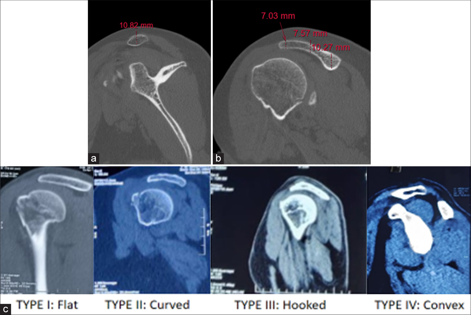
- Parameters from sagittal cuts. (a) Thickness of lateral clavicle (mm), (b) Thickness of acromion (mm), (c) Acromion morphology

- Parameters from axial cuts. Coracoid overlap.
From 3D reconstruction images [Figure 4]: AC joint configuration angles: (A) Acromion angle: Angle between the cranial surface of the acromion and tangent to its articular surface.[9] (B) Clavicle angle: Angle between the cranial surface of the clavicle and tangent to its articular surface.[9] These angles determine the type of AC joint morphology and classify them into:
Overhanging acromion: The acromion angle is more obtuse than the clavicle angle and the difference is more than 10°.
Neutral: Both are acute-angled, or the difference between the two angles is <10°.
Overhanging clavicle: The clavicle angle is more obtuse than the acromion angle and the difference is more than 10°.
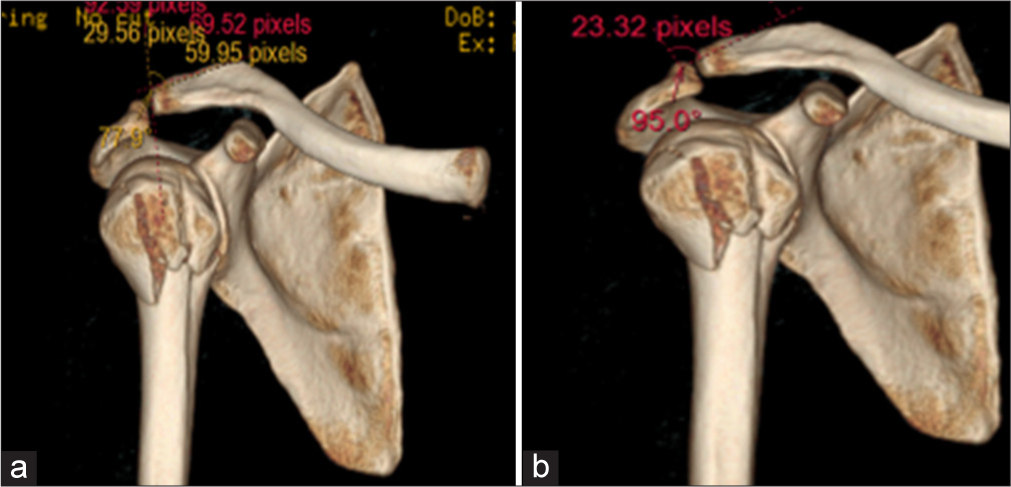
- Parameters from 3D reconstruction images. (a) Clavicular angle (degrees), (b) Acromion angle (degrees).
These parameters were measured, tabulated, and analyzed using statistical analysis to study their variabilities based on age groups and sex. The data entry was done in a Microsoft Excel spreadsheet and the final analysis was done using the Statistical Package for the Social Sciences 25.0 software. For statistical significance, a P < 0.05 was considered statistically significant.
RESULTS
Among the study population, 63 patients belonged to the age group 31–40, followed by 41–50 years with 47, 51–60 years with 41 patients, 61–70 years with 37 patients, 21–30 years with 33, 71–80 years with 24, and >80 years with nine patients. Half of the study population were males (130), and half were females.
The mean value of AC angle, depth of acromion, CC distance, distal clavicle thickness, acromion thickness, CO, acromion angle, and clavicular angle of study subjects are shown in Table 1. In the majority, i.e., 188 (72.3%) patients, acromial morphology was curved, followed by flat (71 [27.3%]). Acromial morphology was hooked in only 1 out of 260 patients (0.4%) [Figure 5]. In most patients (104 = 40%), the AC joint configuration type was an overhanging acromion followed by an overhanging clavicle (90 = 34.7%). AC joint configuration type was neutral in only 66 out of 260 patients (25.4%) [Figure 6]. No significant difference was seen in the depth of the acromion (P = 0.05) between females and males. The mean ± SD of the depth of the acromion in the females was 13.6 ± 2.7, and in the males, it was 14.3 ± 2.6, with no significant difference between them. A significant association was seen in AC angle, CC distance, the thickness of distal clavicle, the thickness of acromion, CO, acromion angle, and clavicular angle with sex (P < 0.05). The mean ± SD of the acromion angle in the females was 98.1 ± 13.4, which was significantly higher than that in the males (92.1 ± 15.9). Mean ± SD of AC angle, CC distance, thickness of distal clavicle, the thickness of acromion, CO, and clavicular angle in males was significantly higher than in females, as outlined in the figure [Figure 7]. There is a notable negative correlation between AC angle and depth of acromion with age, as outlined in Figure 8, hence showing the correspondence between the two factors. The distribution of acromial morphology [Figure 9] was comparable between females and males (Curved: - 76.9% vs. 67.7% respectively, Flat: - 23.1% vs. 31.5% respectively, Hooked: - 0% vs. 0.8% respectively) (P = 0.127).
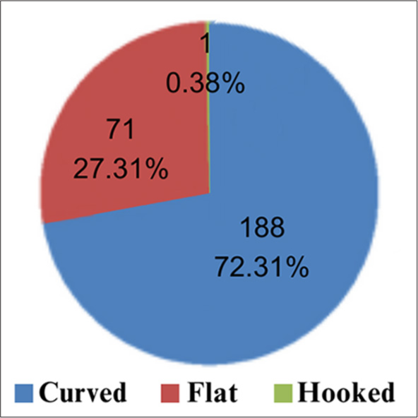
- Distribution of acromial morphology of study subjects.
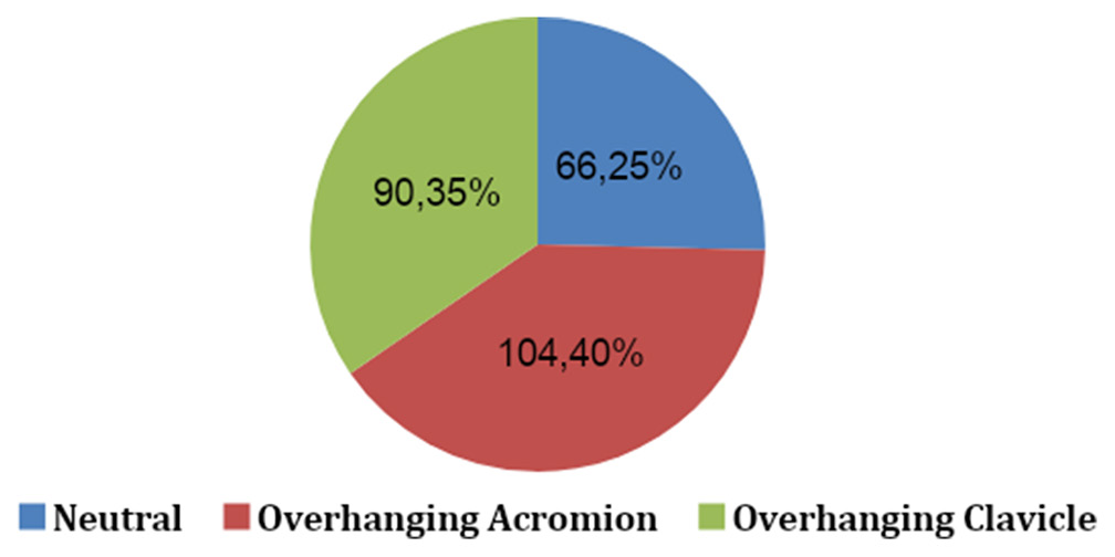
- Distribution of acromioclavicular joint configuration type of study subjects.
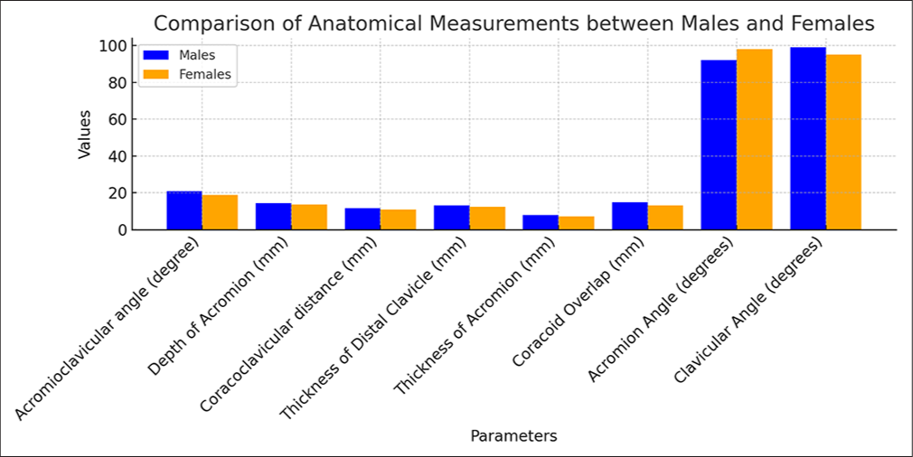
- Association of anatomical parameters with sex. Y-axis: Mean of the variables.

- Correlation of age (years) with acromion depth (mm) and acromioclavicular angle (degrees). Black dots: Individual data of study. Black line: Mean of the data, which shows the negative correlation between acromion depth and the age.
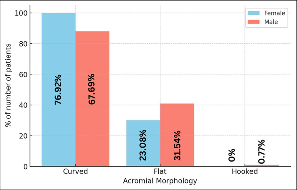
- Association of acromial morphology with sex (Curved: Neutral = 80.30%; Overhanging Acromion = 75%; Overhanging Clavicle = 63.33%; Flat: Neutral = 19.70%; Overhanging Acromion = 24.04%; Overhanging Clavicle = 36.67%; Hooked: Neutral = 0%; Overhanging Acromion = 0.96%; Overhanging Clavicle = 0%).
| Anatomical parameters | Mean±SD | Range |
|---|---|---|
| Acromioclavicular angle (degrees) | 19.78±7.2 | 3.6–44.6 |
| Depth of acromion (mm) | 13.94±2.7 | 5.3–23.12 |
| Coracoclavicular distance (mm) | 11.24±2.56 | 5.46–19.19 |
| Thickness of distal clavicle (mm) | 12.77±2.28 | 7.37–18.8 |
| Thickness of acromion (mm) | 7.53±1.28 | 4.96–12.41 |
| Coracoid overlap (mm) | 14.01±4.18 | 2.33–23.85 |
| Acromion angle (degrees) | 95.13±14.96 | 60.4–128.5 |
| Clavicular angle (degrees) | 97.09±13.56 | 61.6–132.5 |
SD: Standard deviation
A significant negative correlation was seen between age and AC angle, depth of acromion, CC distance, distal clavicle thickness, and acromion thickness, with a correlation coefficient of −0.31, −0.43, −0.47, −0.29, and −0.18, respectively. No correlation was seen between age with CO, acromion angle, and clavicular angle, with correlation coefficients of −0.04, 0.03, and −0.01, respectively [Table 2].
| Variables with correlation to age | Acromioclavicular angle (degrees) | Depth of acromion (mm) |
Coracoclavicular distance (mm) | Thickness of distal clavicle (mm) | Thickness of acromion (mm) | Coracoid overlap (mm) | Acromion angle (degrees) | Clavicular angle (degrees) |
|---|---|---|---|---|---|---|---|---|
| Pearson correlation coefficient | −0.309 | −0.434 | −0.469 | −0.296 | −0.180 | −0.036 | 0.029 | −0.008 |
| P-value | <0.0001 | <0.0001 | <0.0001 | <0.0001 | 0.004 | 0.563 | 0.641 | 0.894 |
DISCUSSION
Many risk factors have been identified in AC joint pathology, the most common being increasing age, especially in degenerative diseases and rotator cuff impingement. As far as AC joint traumatic injuries are considered, various modes of injuries, mechanisms and forces of the trauma determine different patterns of traumatic injury to the AC joint, including AC joint subluxation, dislocations, or fractures. One of the most common ways of surgically stabilizing a high-grade AC joint dislocation or lateral clavicle fracture is a clavicular hook plate, which has its own share of complications associated primarily with a mismatch of the hook to the morphometry of the acromion or AC joint or subacromial space. It becomes important to study the variations in the morphometry of these structures among the population so as to obtain a profile of the subacromial and AC joint morphology. This information can be used to design newer hook plate models that are better contoured to the subacromial space so as to reduce hook plate-related complications such as subacromial impingement, subacromial bursitis, and acromial osteolysis.[10]
In the current study, we evaluated subacromial space morphometry using CT scan-based radiological parameters to analyze and understand these variations in the general population and also to look for sex variabilities in these parameters. According to Azzoni et al.,[11] the size of the subacromial space was related to the acromial morphology, female sex, and rotator cuff pathology.
In the study by Kim et al.,[12] radiological parameters such as AC angle, depth of acromion, AC height difference, clavicular thickness, and acromial thickness were considered for studying population variabilities through CT images. In our study, we have used most of these parameters to study population variations and note sex variations in these entities. According to Garving et al.,[4] rotator cuff tears as well shoulder impingement have their incidence on the rising trend with increasing age, with the peak incidence being in the 5th–6th decade of life.
AC angle is defined as the angle between the clavicle’s upper surface and the acromion’s lower surface. In our study, we attempted to study the variation in this angle among the study population and the pattern of change as age progresses. This angle is directly proportional to the amount of available space in the subacromial space, so it can indirectly predict the subacromial impingement. Kim et al.[12] found the highest variability in these parameters with a mean of 17.1 ± 10.51. In our study population, the mean AC angle was 19.8 ± 7.2, with males having slightly higher values than females. Kim et al.[12] found a slightly higher value among females than males in their study population.
Urist[13] was among the first to study the AC joint morphology and identify three types of AC joint configuration existing in the population under study. Based on this study, we have attempted to evaluate and classify the AC joint configuration among the population under study using CT scan images. According to Crönlein et al.‘s[9] study on a set of 80 healthy populations, in Germany, the most common type was found to be overhanging acromion with 46.2% occurrence, with neutral being second common, followed closely by overhanging clavicle.
In our study, the overhanging acromion was the most common type, with 40% occurrence, followed by the overhanging clavicle, with 34.6% incidence. The neutral type was the least common (25.4%) of AC joint configurations. This finding was consistent with acromial morphology, which showed the curved type to be the most common (72.3%), followed by flat (27.3%) and very rarely hooked (0.4%).
The depth of the acromion showed the extent to which the acromion dipped down below the lateral end of the clavicle. This evaluated the volume of subacromial space. Kim et al.[12] studied this depth among their study population as a probable indicator for the over-reduction of AC joints during surgical stabilization of AC joints. This was also suggested as one of the parameters for matching the hook depth of the hook plate. Depth of the acromion, in our study, did not show any significant sex variations but showed a decreasing trend with increasing age. This suggests that reduced acromial depth is another risk factor for subacromial impingement. The AC angle and acromion depth depend on the integrity of the primary dynamic stabilizers, i.e., the deltoid and trapezius, and the static stabilizers, i.e., the AC and CC ligaments, which explains the variations with age.
CO was measured in the transverse cuts of the CT shoulder. Leite et al.[14] concluded from their study that CO is one of the good predictors of lesions involving subscapularis and long head of biceps lesions. Higher values were suggestive of higher risk. They measured a mean of 14.3 ± 3.9 in a normal population. In our study, we used CO as one of the parameters to study the variability among the study population. The mean CO in our study was found to be about 14.0 ± 4.2. CO has been found to have higher values in the male population, i.e., 14.9 ± 4.7, compared to females, 13.17 ± 3.4.
Acromial and clavicular thickness were considered measurements in the sagittal plane as indicators of morphometry of the AC joint. Elmaraghy et al.,[15] through a cadaveric study of 15 cadavers, noted the mean acromion thickness to be about 5.2 ± 1.4 with significant sex variability in it. The values were found to be larger among males than in females. In our study, mean acromial thickness among the study population was found to be around 7.5 ± 1.3, with a statistically significant higher value among males as compared to females. Lateral clavicle thickness in our study population was found to be 12.8 ± 2.3 on average, with relatively higher values among males than females. As the AC joint and the subacromial space morphometry are influenced by the acromion thickness and lateral clavicle and also as it influences the outcome of surgical fixation of the AC joint at the same time, influences the pattern of traumatic injury or other pathologic occurring at AC joint, the variations in these parameters were studied in the population under study. Kim et al.[12] found the mean acromial and clavicle thickness among the study population to be 8.3 ± 1.1 and 11.7 ± 1.8, with higher values among males than females.
Our study measured CC distance as the vertical distance between the inferior border of the clavicle and the superior border of the coracoid process and is believed to be a measurement of the subacromial space in the coronal plane. Among our study population, we have found a mean value of 11.2 ± 2.6, with males having slightly higher values than females. Nouh et al.[16] studied the AC joint morphometry of 33 patients and 17 patients with evidence of AC joint separations. They determined the normal CC distance to be around 11–13 mm and was found to be influenced by the integrity of CC ligaments. In our study, we have a decreasing pattern in the CC distance, which may be another risk factor in the development of subacromial impingement.
The study has various potential limitations. Further clinical studies or cadaveric studies are required, which can give physical evidence of the relation of these radiographic parameters with the development of subacromial impingement.
CONCLUSION
In this CT-based study, variabilities such as larger AC angle, lower depth of the acromion, reduced CC distance, larger CO, thicker lateral clavicle and acromion, and flat acromion predispose to increased risk of subacromial impingement. This population-based study assesses the range of variabilities that could help develop novel subacromial support devices and augment the existing ones, like hook plates with reduced point force transmission.
Authors’ contributions
TP, MRT, and RDK: Contributed to the conception and design of the study and critical revision of the article. SM: Contributed to the design, data acquisition, statistical analysis, interpretation of the results, and draft manuscript preparation. All Authors have critically reviewed and approved the final draft and are responsible for the manuscript’s content and similarity index.
Ethical approval
The Institutional Ethics Committee, Sri Ramachandra Institute of Higher Education and Research, Chennai, approved the study before data collection (Ref.No.CSP-MED/21/JAN/65/13) on 21/4/2022. The research process and objectives were explained to each participant.
Declaration of patient consent
The authors certify that they have obtained all appropriate patient consent forms. In the form, the patients have given their consent for their images and other clinical information to be reported in the journal. The patients understand that their names and initials will not be published, and due efforts will be made to conceal their identity, but anonymity cannot be guaranteed.
Use of artificial intelligence (AI)-assisted technology for manuscript preparation
The authors confirm that there was no use of artificial intelligence (AI)-assisted technology for assisting in the writing or editing of the manuscript and no images were manipulated using AI.
Conflicts of interest
There are no conflicting relationships or activities.
Financial support and sponsorship: This study did not receive any specific grant from funding agencies in the public, commercial, or not-for-profit sectors.
References
- Shoulder impingement syndrome. Phys Med Rehabil Clin N Am. 2023;34:311-34.
- [CrossRef] [PubMed] [Google Scholar]
- Critical shoulder angle (CSA): Age and gender distribution in the general population. J Orthop Traumatol. 2022;23:10.
- [CrossRef] [PubMed] [Google Scholar]
- Digital technology combined with 3D printing to evaluate the matching performance of ao clavicular hook plates. Indian J Orthop. 2020;54:141-7.
- [CrossRef] [PubMed] [Google Scholar]
- Impingement syndrome of the shoulder. Deutsch Aerztebl Int. 2017;114:765-76.
- [CrossRef] [PubMed] [Google Scholar]
- MRI findings in atraumatic shoulder pain-patterns of disease correlated with age and gender. Ir J Med Sci. 2023;192:847-52.
- [CrossRef] [PubMed] [Google Scholar]
- Morphological characteristics of acromion and acromioclavicular joint in patients with shoulder impingement syndrome and related recommendations: A three-dimensional analysis based on multiplanar reconstruction of computed tomography scans. Orthop Surg. 2021;13:1309-18.
- [CrossRef] [PubMed] [Google Scholar]
- An analysis of acromioclavicular joint morphology as a factor for shoulder impingement syndrome. Shoulder Elbow. 2014;6:165-70.
- [CrossRef] [PubMed] [Google Scholar]
- Coracoid abnormalities and their relationship with glenohumeral deformities in children with obstetric brachial plexus injury. BMC Musculoskelet Disord. 2010;11:237.
- [CrossRef] [PubMed] [Google Scholar]
- Analysis of the bony geometry of the acromio-clavicular joint. Eur J Med Res. 2018;23:50.
- [CrossRef] [PubMed] [Google Scholar]
- Three-dimensional morphometric analysis of the lateral clavicle and acromion: Implications for surgical treatment using subacromial support. SAGE Open Med. 2022;10:20503121221091395.
- [CrossRef] [PubMed] [Google Scholar]
- Sonographic evaluation of subacromial space. Ultrasonics. 2004;42:683-7.
- [CrossRef] [PubMed] [Google Scholar]
- Evaluation of the acromioclavicular joint morphology for minimizing subacromial erosion after surgical fixation of the joint using a clavicular hook plate. Clin Shoulder Elb. 2018;21:138-44.
- [CrossRef] [PubMed] [Google Scholar]
- Complete dislocations of the acromiclavicular joint; The nature of the traumatic lesion and effective methods of treatment with an analysis of forty-one cases. J Bone Joint Surg Am. 1946;28:813-37.
- [Google Scholar]
- Coracohumeral distance and coracoid overlap as predictors of subscapularis and long head of the biceps injuries. J Shoulder Elbow Surg. 2019;28:1723-7.
- [CrossRef] [PubMed] [Google Scholar]
- Subacromial morphometric assessment of the clavicle hook plate. Injury. 2010;41:613-9.
- [CrossRef] [PubMed] [Google Scholar]
- The normal acromioclavicular joint: An in vivo multidetector CT (MDCT) morphometric and biometric cross sectional feasibility study. OMICS J Radiol. 2017;6:260.
- [Google Scholar]


