Translate this page into:
Prevention of lateral plateau fractures during open-wedge high tibial osteotomy: A technical tip and a review of the literature
*Corresponding author: Ahmed M. Ahmed, MRCSEd. Department of Orthopaedic Surgery and Traumatology, Qena Faculty of Medicine, South Valley University, Qena, Egypt. drahmedabdou1993@gmail.com
-
Received: ,
Accepted: ,
How to cite this article: Said E, Addosooki A, Ahmed AM, Tammam H. Prevention of lateral plateau fractures during open-wedge high tibial osteotomy: A technical tip and a review of the literature. J Musculoskelet Surg Res. 2024;8:81-7. doi: 10.25259/JMSR_246_2023
Abstract
Open-wedge high tibial osteotomy is a joint-preserving procedure associated with a number of complications including intra-articular fractures. The primary purpose is to change the varus malalignment into a neutral or valgus alignment according to the extent of cartilage damage. Thus, injury to the lateral tibial plateau would result in serious consequences. This report proposes a simple technical tip to intraoperatively mitigate such a troublesome complication. We also conducted a literature review to investigate the incidence and effects of intra-articular fractures highlighting the techniques recommended by previous authors to avoid intra-articular fractures during tibial valgization osteotomy.
Keywords
Fracture
High tibial osteotomy
Knee
Osteoarthritis
Tibia
INTRODUCTION
High tibial osteotomy (HTO) is a well-known joint-preserving surgery for treating medial compartmental knee osteoarthritis (OA) with varus malalignment. It has gained popularity due to the advantage of joint preservation compared to arthroplasty. The HTO aims to shift the mechanical axis to the relatively healthy lateral knee compartment. It can postpone knee arthroplasty by slowing down the degeneration process.
Debeyre and Artigou were the first to introduce open-wedge HTO (OWHTO) in 1951,[1] and it became widespread in 1987 by Hernigou et al.[2] Several amendments in the surgical techniques have been described, and special implants have been developed.[3] The OWHTO allows maintenance of the bone stock and restoration of normal alignment and does not require fibular osteotomy with an early and active rehabilitation protocol. On the other hand, OWHTO is not free of complications. The previous authors reported a 37–55% complication rate.[4-6] The reported complications included lateral hinge fracture (LHF), delayed wound healing, surgical site infections, vascular injury, venous thromboembolism, alterations in the posterior tibial slope (PTS), overcorrection, undercorrection, delayed union, non-union, implant failure, temporary peroneal nerve paresis, and leg length change.[2,3,7-9]
Intra-articular or lateral tibial plateau (LTP) fractures are the most important complication of OWHTO. It has been reported as early as 1985.[10] Mabrey and McCollum identified 14 complications in 72 knees undergoing OWHTO where the intra-articular fracture was the only complication causing a persistent problem.[11] It is considered a serious adverse event since the lateral compartment of the articular surface is injured. Together with the change of varus malalignment into valgus, a transfer of the weightbearing load from the medial tibial plateau to the injured LTP occurs resulting in poor functional and radiographic outcomes.
The current technical report aimed to describe a simple technical tip that we adopted in our institution to prevent intra-articular fractures during OWHTO and minimize their impact on patients’ outcomes if they occur.
SURGICAL TECHNIQUE
Anteroposterior full-length lower limb weightbearing radiographs are performed preoperatively for deformity analysis. To estimate the desired correction angle, we use the Miniaci et al. planning method considering the associated laxity of the lateral soft tissues measured by the joint line convergence angle to avoid overcorrection.[12]
A 5–8 cm longitudinal skin incision is made extending from the posterior border of the tibial plateau just distal to the joint line to the medial aspect of the tibial tubercle. Then, the superficial part of the medial collateral ligament and pes anserinus is released subperiosteally from the proximal tibia. After that, to expose the osteotomy site and protect the neurovascular bundle, a Hohmann retractor with a blunt tip is positioned beneath the posterior aspect of the upper tibia. Another Hohmann retractor is positioned beneath the patellar tendon.
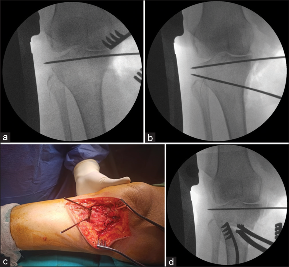
Under fluoroscopic guidance, one or two Kirschner wires (K-wires) are introduced just below and parallel to the articular surface extending from medial to lateral [Figure 1]. The main objective of these subchondral K-wires is to avoid intra-articular extension of the osteotomy during gap distraction. If intra-articular fractures are inevitable, these subchondral wires should maintain the fracture in place until definite osteosynthesis is accomplished.

- (a) Insertion of protective subchondral Kirschner wires (K-wire). (b) Insertion of guide wires along the osteotomy line. (c) Intraoperative image of K-wires inserted during open-wedge high tibial osteotomy. (d) Gradual distraction of the osteotomy gap using a laminar spreader.
Then, two additional parallel K-wires are introduced anteriorly and posteriorly as a guide for the osteotomy plane at a point 40 mm distal to the medial joint line. The guide wires are directed superolaterally, reaching a point 15 mm distal to the lateral joint line [Figure 1]. We utilized the safe zone described by Nakamura et al. located within and lateral to the medial margin of the proximal tibiofibular junction (PTFJ) for the hinge point.[13]
An oscillating saw is used to perform the horizontal osteotomy distal to the guidewires extending to 10 mm medial to the lateral cortex. A second complete vertical osteotomy is performed with a thinner blade starting from the proximal to the insertion of the patellar tendon and directed at an angle of 120° to the horizontal osteotomy plane.
Then, we hammer step-by-step chisels of increasing widths into the osteotomy gap. Subsequently, a gradual distraction of the osteotomy is carried out using a bone spreader. After achieving the desired degree of correction, the leg is extended with the patella facing forward. A laminar spreader is inserted in the posterior part of the osteotomy gap to maintain the achieved opening distance while preserving the PTS [Figure 1]. After a desired angular correction is achieved, internal fixation is carried out.
Isometric exercises and straight leg raises are initiated in the early postoperative period. Partial weightbearing should be allowed at three weeks if there is radiographic evidence of gap healing. Full weightbearing should be started at six weeks postoperatively.
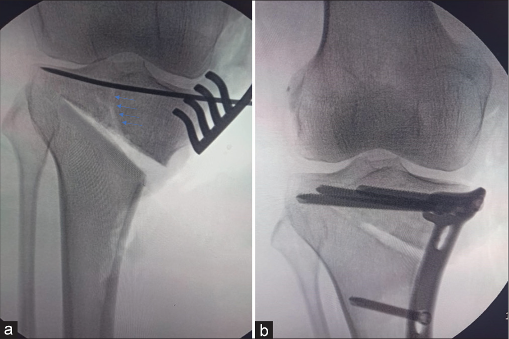
A 42-year-old female patient presented with symptomatic knee varus deformity and OA of the medial compartment treated with OWHTO and fixed by the TomoFix locking plate (OrthoMed, Egypt) [Figure 2]. Intraoperatively, a fracture line was noted extending from the osteotomy plane into the articular surface of the tibial head during the gap opening. The preliminary subchondral K-wire maintained the plateau fracture in place until definite fixation using the same plate was done. At six weeks postoperatively, the union was achieved. Full weightbearing was permitted after eight weeks with no subsequent complications.

- (a) Intra-articular fracture (blue arrows) during gap opening where displacement was prevented by the subchondral Kirschner wires. (b) Definite fixation of intra-articular fracture during high tibial osteotomy using the TomoFix plate.
DISCUSSION
The HTO is a technically challenging procedure that might be complicated by intra- and extra-articular fractures. Extra-articular fractures (classified as LHFs type I and II) have no significant impact on radiologic or functional outcomes.[14] On the contrary, intra-articular or LTP fractures (sometimes classified as LHF type III) are unstable. They may result in union problems and correction loss or alter the PTS.[15] From a biomechanical point of view, type I LHFs represent the propagation of the osteotomy line within the PTFJ, which is surrounded by dense connective tissue. Its stability is due to the compressive forces exerted by weightbearing while the fibula supports the LTP.
On the other hand, type II LHFs involve the distal part of the PTFJ where the weightbearing axis is transferred to the fibula. In type III LHFs, the proximal segment seems to be only supported by the implant.[16]
Although most intra-articular fractures occur intraoperatively, they may not be easily detectable on conventional radiographs.
Therefore, computed tomography may be necessary for early detection.[17] Tibial head infraction starts in the cancellous bone, which can be discovered by continuous radiographic control. To minimize the amount of radiation exposure intraoperatively, Lee et al. described the unequal separation of proximal and distal segments during the gap-opening process as an early sign of LTP fractures. The sign is attributed to the medial shift of the pivoting center from the lateral hinge to the lateral plateau.[18]
Several studies reporting different rates of intra-articular fractures during OWHTO over the past two decades [Table 1]. The overall rate of intraoperative LTP fractures ranged from 1% to 19%. A recent study by Jin et al. of 339 knees reported intra-articular fractures as the most common complication in their series.[19] Some authors described intra-articular fractures as a minor complication with no significant effect on surgical outcomes or rehabilitation programs. Others reported the need for additional fixation by screws or plates, revision surgery, delayed union, alignment overcorrection, undercorrection, loss of correction, limited range of motion, and delayed weightbearing.
| First author | Year | No. knees | Implant | No. LTP fractures (%) |
Follow-up | Comment |
|---|---|---|---|---|---|---|
| Spahn[22] | 2003 | 55 30 |
Puddu C-plate | 8 (14.6) 2 (6.6) |
- | Need a higher correction of 11° Additional osteosynthesis |
| Amendola[26] | 2004 | 74 | Puddu | 7 (19) | 16–26 m | - |
| Esenkaya and Elmali[27] |
2006 | 58 | 2- or 4-hole wedge plate |
5 (8.6) | 21 m | Limited ROM to 70° on discharge |
| Asik[28] | 2006 | 65 | Puddu | 1 (1.5) | 34 m | Additional osteosynthesis |
| Niemeyer[29] | 2007 | 43 | TomoFix | 1 (2.3) | Min 2 year | Revision surgery using screws on 3rd post-operative day |
| Niemeyer[30] | 2010 | 69 | TomoFix | 1 (1.4) | Min 36 m | Additional osteosynthesis |
| Nelissen[5] | 2010 | 49 | Puddu | 3 (6.1) | - | - |
| Song[21] | 2010 | 90 | Aescula | 6 (6.7) | 26.7 m | Additional osteosynthesis or casting Healed at 3 m |
| Takeuchi[15] | 2010 | 27 | TomoFix | 1 (3.7) | 61 m | Insignificant effect |
| Takeuchi[15] | 2011 | 104 | TomoFix | 2 (2) | 41 m | Insignificant effect |
| de Mello Junior[31] | 2011 | 67 | Puddu | 2 (4.4) | Min 12 m | - |
| Chae[32] | 2011 | 138 | Locking T-plate | 3 (2.2) | 36.8 m | Additional osteosynthesis (plate) |
| Schröter[33] | 2011 | 35 | Position HTO plate |
1 (3) | Min 12 m | Additional osteosynthesis (screws) |
| Saragaglia[34] | 2011 | 124 | AO T-plate | 9 (7.25) | 10.39 year | Insignificant effect |
| Tabrizi[35] | 2013 | 21 | L-plate T-plate |
2 (12.5) | 6 m | Insignificant effect |
| Jung[18] | 2013 | 92 94 |
TomoFix Aescula |
3 (3) 2 (2) |
2 year | - |
| Martin[36] | 2014 | 323 | TomoFix Puddu |
9 (2.8) | 39.5 m | Delayed weight-bearing |
| Giuseffi[37] | 2015 | 89 | Arthrex wedge plate |
6 (6.7) | 4 year | Insignificant effect |
| Nakamura[16] | 2015 | 47 | TomoFix | 6 (12.8) | Min 1 year | Additional osteosynthesis Low intensity ultrasound pulse Delayed union Overcorrection and loss of correction |
| Nakamura[13] | 2017 | 111 | TomoFix | 8 (7.2) | - | Additional osteosynthesis (screws) Revision for undercorrection Overcorrection and loss of correction |
| Ogawa[20] | 2017 | 82 | TomoFix | 4 (4.9) | 20.2 m | - |
| Han[38] | 2019 | 209 | TomoFix | 2 (1) | Min 2 year | Insignificant effect |
| Tuhanioglu[39] | 2019 | 18 | Locked wedge HTO plates |
1 (5.6) | 31.61 m | - |
| Hartz[40] | 2019 | 346 | PEEK power HTO plate |
7 (2) | Min 1 year | Insignificant effect |
| Schwartz[41] | 2019 | 19 | 4-hole plate | 1 (5.3) | Min 1 year | Partial loss of correction Delayed union |
| Yabuuchi[42] | 2020 | 85 | TomoFix | 4 (4.7) | 4.5 year | Loss of correction>5° Change rehabilitation protocol |
| Sidhu[43] | 2020 | 200 | TomoFix | 6 (3) | Min 2 year | Delayed union Delayed weight-bearing |
| Jin[19] | 2020 | 339 | Aescula | 12 (3.5) | 9.6 year | Insignificant effect |
OWHTO: Open-wedge high tibial osteotomy, HTO: High tibial osteotomy, LTP: Lateral tibial plateau, PEEK: Polyetheretherketone, ROM: Range of motion
Intraoperative LTP fractures usually arise secondary to technical problems and inadequate surgical experience. The previous studies demonstrated several factors contributing to the propagation of the osteotomy to the articular surface such as incomplete anterior or posterior corticotomy, high-level osteotomy above the fibular head closer to the LTP joint line, and application of excessive valgus force during the gap opening. Furthermore, using thick osteotomes, large correction angles (>−12°), and the creation of inappropriate osteotomies not parallel to the PTS make intra-articular fractures more likely to occur.[20-22]
In Table 2, we summarized several technical notes recommended by previous authors in terms of direction, depth, creation, and opening of the osteotomy so that knee surgeons would avoid such troublesome complications. Recently, novel devices have been developed to decrease the rate of intraoperative complications such as LTP fractures. Akamatsu et al. showed that navigation systems provided more satisfactory radiographic outcomes in the context of LTP fractures compared to conventional techniques.[23] Based on the fact that intra-articular fracture is attributed to the misplacement of the osteotomy bone cut and using a spacer for osteotomy gap opening, Ribeiro et al. performed OWHTO on eight cadaveric knees using a realignment high control system.[24] The system consisted of an implant linked to a dynamic instrument so that the plate could be fixed before opening the osteotomy gap. This allowed accurate mechanical correction and appropriate control of the PTS. Furthermore, Ghinelli et al. examined the iBalance HTO system, which consists of a non-absorbable polyetheretherketone implant and anchors inserted into the opening wedge osteotomy site.[25] Although no intra-articular fractures were reported in either technique, the cost and unavailability of such complex devices hinder their routine application.
| Surgical step | First author, year | Recommendation |
|---|---|---|
| Direction of osteotomy | Amendola, 2004[26] | The osteotomy should be more horizontal, away from the joint line>1.5 cm. |
| Chae, 2011[32] | The osteotomy should be directed toward a “safe zone” around 1.6 cm (range, 1.2-1.8) and extending from 0.8 cm distal to the lateral tibial joint line to the fibular circumference line. | |
| Vanadurongwan, 2013[44] | The osteotomy should be directed toward the “anatomical safe zone” between articular cartilage of posterolateral proximal tibia and PTFJ. | |
| Nakamura, 2017[13] | The osteotomy should be directed toward zone WL (within the PTFJ, lateral to the medial margin of the PTFJ). The bone density at the level of the PTFJ is higher than that above or below it and the PTFJ has many soft-tissue insertions which can confer stability even if the osteotomy direction or force is suboptimal. | |
| Depth of osteotomy | Jacobi, 2010[45] and Schwartz, 2018[41] | The saw must cut until it is going through the external cortex to achieve an almost complete osteotomy with temporary external fixation of the lateral hinge. |
| Creation of osteotomy | Amendola, 2004[26] | Thin AO osteotomes are preferred. |
| Jacobi, 2010[45] | Osteotomy is done below a K-wire which guides the saw blade and osteotomy chisels. | |
| Opening of osteotomy | Esenkaya and Elmali, 2006[27] and Lee, 2013[18] | Enlargement of the osteotomy must be performed very slowly and cautiously below the guide wires. After sawing, chisels of increasing widths must be hammered step by step into the osteotomy space, followed by a gradual controlled opening through the plastic deformation zone (a spreader-chisel or osteotomy jack could be used). A lever force must be strictly avoided. |
OWHTO: Open-wedge high tibial osteotomy, PTFJ: Proximal tibiofibular junction, K-wires: Kirschner wires, AO: Arbeitsgemeinschaft für Osteosynthesefragen,
CONCLUSION
In the current technical report, we described a quite simple and applicable technique using readily available tools to mitigate intra-articular fractures during HTO. Insertion of subchondral K-wires before implementation of the desired osteotomy will not only decrease the risk of intraoperative LTP fractures but also keep the intra-articular fracture, if it occurs, undisplaced until definite fixation is achieved. Nevertheless, a large-scale case series is necessary to support our conclusion.
AUTHORS’ CONTRIBUTIONS
ES and AA carried out the conception and technique idea and performed the surgery. AMA and HT carried out data acquisition and performed the literature search. All authors drafted the manuscript and critically reviewed and approved the final draft, and responsible for the manuscript’s content and similarity index.
ETHICAL APPROVAL
The Institutional Review Board approval is not required.
DECLARATION OF PATIENT CONSENT
The authors certify that they have obtained all appropriate patient consent forms. In the form, the patient has given her consent for her images and other clinical information to be reported in the journal. The patient understands that her name and initials will not be published, and due efforts will be made to conceal her identity, but anonymity cannot be guaranteed.
USE OF ARTIFICIAL INTELLIGENCE (AI)-ASSISTED TECHNOLOGY FOR MANUSCRIPT PREPARATION
The authors confirm that there was no use of artificial intelligence (AI)-assisted technology for assisting in the writing or editing of the manuscript and no images were manipulated using AI.
CONFLICTS OF INTEREST
There are no conflicting relationships or activities.
FINANCIAL SUPPORT AND SPONSORSHIP
This study did not receive any specific grant from funding agencies in the public, commercial, or not-for-profit sectors.
References
- Long term results of 260 tibial osteotomies for frontal deviations of the knee. Rev Chir Orthop Reparatrice Appar Mot. 1972;58:335-9.
- [Google Scholar]
- Proximal tibial osteotomy for osteoarthritis with varus deformity. A ten to thirteen-year follow-up study. J Bone Joint Surg Am. 1987;69:332-54.
- [CrossRef] [PubMed] [Google Scholar]
- Improvements in surgical technique of valgus high tibial osteotomy. Knee Surg Sports Traumatol Arthrosc. 2003;11:132-8.
- [CrossRef] [PubMed] [Google Scholar]
- The effect of lateral cortex disruption and repair on the stability of the medial opening wedge high tibial osteotomy. Am J Sports Med. 2005;33:1552-7.
- [CrossRef] [PubMed] [Google Scholar]
- Stability of medial opening wedge high tibial osteotomy: A failure analysis. Int Orthop. 2010;34:217-23.
- [CrossRef] [PubMed] [Google Scholar]
- Evolution of open-wedge high-tibial osteotomy: Experience with a special angular stable device for internal fixation without interposition material. Int Orthop. 2010;34:167-72.
- [CrossRef] [PubMed] [Google Scholar]
- Open wedge tibial osteotomy with acrylic bone cement as bone substitute. Knee. 2001;8:103-10.
- [CrossRef] [PubMed] [Google Scholar]
- Medial opening-wedge high tibial osteotomy with use of porous hydroxyapatite to treat medial compartment osteoarthritis of the knee. J Bone Joint Surg Am. 2003;85:78-85.
- [CrossRef] [PubMed] [Google Scholar]
- Lateral tibial plateau fracture as a postoperative complication of high tibial osteotomy. Orthopedics. 1985;8:1009-13.
- [CrossRef] [PubMed] [Google Scholar]
- High tibial osteotomy: A retrospective review of 72 cases. South Med J. 1987;80:975-80.
- [CrossRef] [PubMed] [Google Scholar]
- Proximal tibial osteotomy. A new fixation device. Clin Orthop Relat Res. 1989;246:250-9.
- [CrossRef] [Google Scholar]
- Appropriate hinge position for prevention of unstable lateral hinge fracture in open wedge high tibial osteotomy. Bone Joint J. 2017;99b:1313-8.
- [CrossRef] [PubMed] [Google Scholar]
- Extra-articular lateral hinge fracture does not affect the outcomes in medial open-wedge high tibial osteotomy using a locked plate system. Arthroscopy. 2018;34:3246-55.
- [CrossRef] [Google Scholar]
- Fractures around the lateral cortical hinge after a medial opening-wedge high tibial osteotomy: A new classification of lateral hinge fracture. Arthroscopy. 2012;28:85-94.
- [CrossRef] [PubMed] [Google Scholar]
- The validity of the classification for lateral hinge fractures in open wedge high tibial osteotomy. Bone Joint J. 2015;97B:1226-31.
- [CrossRef] [PubMed] [Google Scholar]
- Hinge fractures are underestimated on plain radiographs after open wedge proximal tibial osteotomy: Evaluation by computed tomography. Am J Sports Med. 2019;47:1370-5.
- [CrossRef] [PubMed] [Google Scholar]
- An early sign of intraarticular fracture of the lateral tibial plateau during opening wedge high tibial osteotomy. Knee. 2013;20:66-8.
- [CrossRef] [PubMed] [Google Scholar]
- Survival and risk factor analysis of medial open wedge high tibial osteotomy for unicompartment knee osteoarthritis. Arthroscopy. 2020;36:535-43.
- [CrossRef] [PubMed] [Google Scholar]
- The prevention of a lateral hinge fracture as a complication of a medial opening wedge high tibial osteotomy: A case control study. Bone Joint J. 2017;99B:887-93.
- [CrossRef] [PubMed] [Google Scholar]
- The complications of high tibial osteotomy: Closing-versus opening-wedge methods. J Bone Joint Surg Br. 2010;92:1245-52.
- [CrossRef] [PubMed] [Google Scholar]
- Complications in high tibial (medial opening wedge) osteotomy. Arch Orthop Trauma Surg. 2004;124:649-53.
- [CrossRef] [PubMed] [Google Scholar]
- Navigated opening wedge high tibial osteotomy improves intraoperative correction angle compared with conventional method. Knee Surg Sports Traumatol Arthrosc. 2012;20:586-93.
- [CrossRef] [PubMed] [Google Scholar]
- A novel device for greater precision and safety in open-wedge high tibial osteotomy: Cadaveric study. Arch Orthop Trauma Surg. 2020;140:203-8.
- [CrossRef] [PubMed] [Google Scholar]
- High tibial osteotomy for the treatment of medial osteoarthritis of the knee with new iBalance system: 2 years of follow-up. Eur J Orthop Surg Traumatol. 2016;26:523-35.
- [CrossRef] [PubMed] [Google Scholar]
- Opening wedge high tibial osteotomy using a novel technique: Early results and complications. J Knee Surg. 2004;17:164-9.
- [CrossRef] [PubMed] [Google Scholar]
- Proximal tibia medial open-wedge osteotomy using plates with wedges: Early results in 58 cases. Knee Surg Sports Traumatol Arthrosc. 2006;14:955-61.
- [CrossRef] [PubMed] [Google Scholar]
- High tibial osteotomy with Puddu plate for the treatment of varus gonarthrosis. Knee Surg Sports Traumatol Arthrosc. 2006;14:948-54.
- [CrossRef] [PubMed] [Google Scholar]
- Two-year results of open-wedge high tibial osteotomy with fixation by medial plate fixator for medial compartment arthritis with varus malalignment of the knee. Arthroscopy. 2008;24:796-804.
- [CrossRef] [PubMed] [Google Scholar]
- Open-wedge osteotomy using an internal plate fixator in patients with medial-compartment gonarthritis and varus malalignment: 3-year results with regard to preoperative arthroscopic and radiographic findings. Arthroscopy2010;. ;26:1607-16.
- [CrossRef] [PubMed] [Google Scholar]
- Complications following medial opening wedge osteotomy of the knee: Retrospective study. Rev Brasil Ortop. 2011;46:64-8.
- [CrossRef] [PubMed] [Google Scholar]
- Early complications of medial opening wedge high tibial osteotomy using autologous tricortical iliac bone graft and T-plate fixation. Knee. 2011;18:278-84.
- [CrossRef] [PubMed] [Google Scholar]
- High complication rate after biplanar open wedge high tibial osteotomy stabilized with a new spacer plate (Position HTO plate) without bone substitute. Arthroscopy. 2011;27:644-52.
- [CrossRef] [PubMed] [Google Scholar]
- Outcome of opening wedge high tibial osteotomy augmented with a Biosorb® wedge and fixed with a plate and screws in 124 patients with a mean of ten years follow-up. Int Orthop. 2011;35:1151-6.
- [CrossRef] [PubMed] [Google Scholar]
- A short term follow up comparison of genu varum corrective surgery using open and closed wedge high tibial osteotomy. Malays Orthop J. 2013;7:7-12.
- [CrossRef] [PubMed] [Google Scholar]
- Adverse event rates and classifications in medial opening wedge high tibial osteotomy. Am J Sports Med. 2014;42:1118-26.
- [CrossRef] [PubMed] [Google Scholar]
- Opening-wedge high tibial osteotomy: Review of 100 consecutive cases. Arthroscopy. 2015;31:2128-37.
- [CrossRef] [PubMed] [Google Scholar]
- Complications associated with medial opening-wedge high tibial osteotomy using a locking plate: A multicenter study. J Arthroplasty. 2019;34:439-45.
- [CrossRef] [PubMed] [Google Scholar]
- High tibial osteotomy in obese patients: Is successful surgery enough for a good outcome? J Clin Orthop Trauma. 2019;10(Suppl 1):S168-73.
- [CrossRef] [PubMed] [Google Scholar]
- Plate-related results of opening wedge high tibial osteotomy with a carbon fiber reinforced poly-ether-ether-ketone (CF-PEEK) plate fixation: A retrospective case series of 346 knees. J Orthop Surg Res. 2019;14:466.
- [CrossRef] [PubMed] [Google Scholar]
- Minimally invasive opening wedge tibia outpatient osteotomy, using screw-to-plate locking technique and a calcium phosphate cement. Eur J Orthop Surg Traumatol. 2018;28:799-809.
- [CrossRef] [PubMed] [Google Scholar]
- Clinical outcomes and complications during and after medial open-wedge high tibial osteotomy using a locking plate: A 3-to 7-year follow-up study. Orthop J Sports Med. 2020;8:2325967120922535.
- [CrossRef] [PubMed] [Google Scholar]
- Low rates of serious complications but high rates of hardware removal after high tibial osteotomy with Tomofix locking plate. Knee Surg Sports Traumatol Arthrosc. 2021;29:3361-7.
- [CrossRef] [PubMed] [Google Scholar]
- The anatomical safe zone for medial opening oblique wedge high tibial osteotomy. Singapore Med J. 2013;54:102-4.
- [CrossRef] [PubMed] [Google Scholar]
- Avoiding intraoperative complications in open-wedge high tibial valgus osteotomy: Technical advancement. Knee Surg Sports Traumatol Arthrosc. 2010;18:200-3.
- [CrossRef] [PubMed] [Google Scholar]


