Translate this page into:
Case of traumatic ankle pain
Corresponding Author:
Nizar A Al-Nakshabandi
Department of Radiology, College of Medicine, King Saud University, Riyadh
Saudi Arabia
nizar97@hotmail.com
| How to cite this article: Al-Nakshabandi NA. Case of traumatic ankle pain. J Musculoskelet Surg Res 2020;4:237-238 |
History
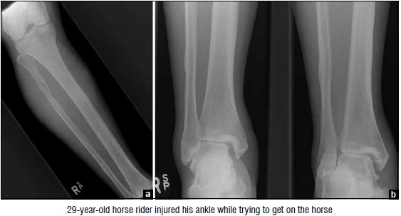
A 29-year-old horse rider injured his ankle while trying to get on the horse. He fell with the foot and leg hitting the ground at an awkward angle while he was rotating. He presented with ankle pain, swelling, bruising, and restricted range of motion.
Findings
The anteroposterior (AP) view of the tibia and fibula (a) demonstrates a spiral fracture of the proximal fibula. Furthermore, the ankle AP, and oblique views (b and c, respectively) demonstrate the widening of the ankle joint due to distal tibiofibular syndesmosis or deltoid ligament disruption. The talus is not sitting symmetrically in the ankle mortise.
Diagnosis
Maisonneuve-fracture dislocation.
Pearls and Discussion
The mechanism of injury in these fracture-dislocation is pronation with external rotation mechanism. Classically in the examination situation, you would be given and AP and oblique view of the ankle and once you detect the tibiofibular syndesmosis disruption or suspected deltoid ligament injury, the examiner will tell you would you like to see any other images or order any other views you should mention that you would like to take a look at the proximal tibia and fibula where you will see the spiral fracture of the fibula. Alternatively, you may just be given the AP view of the proximal tibia and fibula and you should then ask for the ankle views.
The proximal fibular fracture does not require any surgical treatment; however, the syndesmosis will require approximation and fixation. The use of one screw versus two screws and three cortices versus four are all controversial. The most critical aspect is the anatomical reduction of the distal fibula to the tibia, maintaining the normal anatomical relationship.
Declaration of patient consent
The author certifies that he has obtained all appropriate patient consent forms. In the form, the patient has given his consent for his images and other clinical information to be reported in the journal. The patient understands that his name and initials will not be published and due efforts will be made to conceal his identity, but anonymity cannot be guaranteed.
Financial support and sponsorship
This study did not receive any specific grant from funding agencies in the public, commercial, or not-for-profit sectors.
Conflicts of interest
There are no conflicts of interest.
Further reading
- Forster BB, Lee JS, Kelly S, O′Dowd M, Munk PL, Andrews G, et al. Proximal tibiofibular joint: An often-forgotten cause of lateral knee pain. AJR Am J Roentgenol 2007;188:W359?66.
- Maisonneuve JG. Research of Fibula Fractures. Paris, France: Loquin & Cie; 1840.
Fulltext Views
1,350
PDF downloads
1,213


