
Translate this page into:
Management of closed diaphyseal fractures in children in two black African countries: Preliminary results and epidemiological profile
*Corresponding author: Hermann Victoire Feigoudozoui, Department of Orthopaedic and Trauma Surgery, Félix Houphouët-Boigny University, Abidjan, Cote D’Ivoire. hfeigoudozoui@gmail.com
-
Received: ,
Accepted: ,
How to cite this article: Feigoudozoui H, Mapouka M, Parteina D, Moun-Goss N, N’doma Ngatchoukpo V, Bankolé S. Management of closed diaphyseal fractures in children in two black African countries: Preliminary results and epidemiological profile. J Musculoskelet Surg Res. 2024;8:221-6. doi: 10.25259/JMSR_93_2024
Abstract
Objectives:
The objective of this study was to describe the clinical evolution and the epidemiological profile of long bone fractures in children in two African countries.
Methods:
This was a five-year retrospective study of closed fractures of long bones in children aged 9.7 years (Ranging 4–16). Seventy-two fractures were treated by elastic stable intramedullary nailing and plate osteosynthesis. The results were evaluated according to Oestern and Tscherne criteria, and parental satisfaction was assessed using an individual investigation form.
Results:
The treatment of closed fractures of long bones in children is marked by complications such as infection (16.6%), implant migration (19.4%), non-union (11.1%), and malunion (2.7%). According to the assessment criteria, 33.3% were excellent, 45.8% were good, and 20.8% were poor. Twenty percentages of parents were very satisfied, 61% were satisfied, and 18% were disappointed with the outcome.
Conclusion:
Osteosynthesis is only indicated in common pediatric trauma if the capabilities of remodeling a callous are insufficient. It should not be the first choice in children’s fracture therapeutic options.
Keywords
Africa
Children
Fracture
Long bone
Osteosynthesis

INTRODUCTION
More than 30% of children suffer at least one fracture before the age of 17.[1] Open or closed fractures of the long bones in young children are not uncommon. Closed stable fractures are preferably treated conservatively.[2-4] Unstable fractures are usually treated surgically.[5] The most widely used osteosynthesis for diaphyseal fractures of the long bones in children is elastic stable intramedullary nailing (ESIN).[4,6] Surgical treatment is often associated with complications, the most common of which are infection, pseudoarthrosis, and malunion.[6-8]
The risk of post-operative complications is increased in poor conditions in developing countries. The literature on the surgical treatment of long bones in developing countries (Africa, Asia, and Latin America) is poor. The importance of this study was to enrich the knowledge around this subject. Finally, this study describes the realities of surgical treatment of long bones in low-income countries, taking a typical example.
This study aimed to describe the clinical course of osteosynthesis and the epidemiological profile of closed diaphyseal fractures in children in developing countries.
MATERIALS AND METHODS
Between January 2017 and December 2021, in the Ivory Coast and Central African Republic, a single-center retrospective study was conducted on 89 children with a mean age of 9.7 years (Ranging 4–16). Seventeen children were lost to follow-up. Seventy-two cases were collected, including 41 boys and 31 girls, with a sex ratio of 4:1. Open fractures, pathological fractures, children under four years of age, and conservatively treated fractures were excluded. Children over six years of age were the majority (64%). The circumstances of occurrence were predominantly play accidents (62%). The right side was the dominant side (68%). There was no bilaterality. The most common fractures were those of the femur, humerus, and tibia. The average time to treatment was 4.7 days (Ranging 1–62). Treatment by ESIN or plate osteosynthesis was indicated either immediately or after the failure of conservative treatment. Most of the indications were for closed ESIN. More than half of the fractures had healed. The minimum follow-up was 24 months. The criteria of Oestern and Tscherne[2] were used to assess clinical and functional outcomes [Table 1]. Parental satisfaction was obtained through an individual form. The data were processed and analyzed using Epi Info software. Statistical tests were performed using Pearson’s Chi-square test, with a significance threshold called P < 5% (<0.05).
| Result | Joint mobility deficiency | Function | Complaints |
|---|---|---|---|
| Excellent | Elbow | No limitation of strength or nerve function | None |
| Extension 10 | |||
| Flexion 15 | |||
| Wrist | |||
| Pronation/Supination 15 | |||
| Flexion (dorsal/volar) 10 | |||
| Abduction (radial/ulnar) 2 | |||
| Good | Elbow | - Change of normal activity for the child | Subjective minimal complaints |
| Extension 10 | - Choice of sports activities | without joint mobility deficit | |
| Flexion 30 | |||
| Wrist | |||
| Pronation/Supination 25 | |||
| Flexion (dorsal/volar) 25 | |||
| Abduction (radial/ulnar) 10 | |||
| Average | Elbow | -Mild-to-moderate strength deficit | Greater subjective |
| Extension 20 | -Impairment of pre-operative nerve function | complaints with all movements | |
| Flexion 45 | |||
| Wrist | |||
| Pronation/Supination 45 | |||
| Flexion (dorsal/volar) 45 | |||
| Abduction (radial/ulnar) 10 | |||
| Poor | All deficits greater than the previous ones | - Very sharp drop in strength | Substantial subjective complaints with reduced joint mobility |
| - Nerve function impairment without pre-operative lesion |
RESULTS
At a minimal follow-up of 24 months, 72 fractures had been treated using ESIN or plate osteosynthesis and screws, 64 of which were healed [Table 2]. Twenty-six (36.1%) fractures were treated by plate osteosynthesis, including 23 (31.9%) on the femur and three (4.1%) on the tibia. Fourteen (19.4%) fractures were treated by ESIN, including 23 (31.9%) on the humerus, 9 (12.5%) on the forearm, 3 (4.1%) on the femur, and 11 (15.2%) on the tibia. Thirty-six (50%) fixations developed complications [Table 3]. Statistical analysis demonstrated a relationship between the various bones and complications. We found 12 (16.6%) cases of surgical site infection, including 5 (6.9%) on the femur and 7 (9.7%) on the tibia. There were 14 (19.4%) cases of implant migration, including 4 (5.5%) to the humerus, 5 (6.9%) to the forearm, and 5 (6.9%) to the femur. There were 8 (11.1%) cases of non-union, including 2 (2.7%) on the humerus, 4 (5.5%) on the femur, and 2 (2.7%) on the tibia. We also found 2 (2.7%) cases of malunion on the femur and tibia.
| Data | n(%) |
|---|---|
| Age | |
| <6 years | 8 (11.1) |
| >6 years | 64 (88.8) |
| Sex | |
| Female | 31 (43) |
| Male | 41 (56.9) |
| Circumstance of occurrence | |
| Playground accident | 45 (62.5) |
| Road traffic accident | 27 (37.5) |
| Side | |
| Right side | 49 (68) |
| Left side | 23 (31.9) |
| The bone | |
| Humerus | 23 (31.9) |
| Forearm | 9 (12.5) |
| Femur | 26 (36.1) |
| Tibia | 14 (19.4) |
| Consolidation | |
| Union | 64 (88.8) |
| Non-union | 8 (11.1) |
| Items | Humerus | Forearm | Femur | Tibia | Total |
|---|---|---|---|---|---|
| Implant | |||||
| Plate osteosynthesis | - | - | 23 | 3 | 26 |
| ESIN | 23 | 9 | 3 | 11 | 14 |
| Total | 23 | 9 | 26 | 14 | 72 |
| Complication | |||||
| Infection | - | - | 5 | 7 | 12 |
| Implant migration | 4 | 5 | 5 | - | 14 |
| Non-union | 2 | - | 4 | 2 | 8 |
| Malunion | - | - | 1 | 1 | 2 |
| Total | 6 | 5 | 15 | 10 | 36 |
ESIN: Elastic stable intramedullary nail
The relationship between surgical indications and postoperative complications was proven with a significant probability (P < 0.5). We found that of the 13 (18%) cases of surgical site infection, 11 (15.2%) followed plate osteosynthesis. Among the 13 (18%) cases of implant migration, the most frequent was ESIN in 13 (18%) cases. The 8 (11.1%) cases of non-union all occurred following plate osteosynthesis. Finally, of the 3 (4.1%) cases of malunion, 2 (2.7%) followed ESIN.
According to the Oestern and Tscherne evaluation criteria, 24 (33.3%) outcomes were excellent, 33 (45.8%) were good, and 15 (20.8%) were poor. The Oestern and Tscherne criteria are a set of scores for the functional assessment of long-bone fractures in children. It is an assessment score based on joint mobility, limb function, patient complaints, bone consolidation, and bone mobility.
Finally, 15 (20.8%) parents were very satisfied, 44 (61.1%) were satisfied, and 13 (18%) were disappointed with the outcome. The mean post-operative hospital stay was 11.3 days (Ranging 2–32).
DISCUSSION
Authors generally believe conservative treatment is the treatment of choice for fractures in children.[2-4] However, in this study, the problem concerned diaphyseal fractures. Conservative treatment is sometimes difficult to achieve due to genuine instability or irreducibility. This justifies the option of surgery. Osteosynthesis, thus, has its place in the treatment of fractures in children. It must be minimally invasive, respecting the rules of osteosynthesis and periosteal consolidation in children.
The predominant proportions concerning male gender, age over ten years, and play injury are similar to those of other studies.[2,4,9,10] Boys are more mobile and active than girls and are exposed to accidents of all kinds. The age group over 10 is the most mobile and reckless in childhood and, therefore, is exposed to accidents in everyday activities. The dominant side has an interest in the occurrence of complications.
Using ESIN for osteosynthesis in children is frequent and less invasive for the growth plate. We believe that it remains the standard technique for treating diaphyseal fractures in children. Of the fractures treated with ESIN, 17 had postoperative complications [Figures 1-3]. There were two cases of infection, 13 cases of K-wire migration, and three cases of malunion. The K-wire migration was always caused by early weight-bearing. The fractures that healed into a malunion were medio-diaphyseal transverse fractures. These complications were linked to the parent’s failure to follow post-operative instructions. In addition to infection (three cases), Yaokreh et al. found three cases of skin irritation and two cases of joint stiffness in 26 fractures treated by ESIN.[9] In their opinion, these three cases of infection were superficial infections caused by insufficient skin disinfection and longer operating times. However, the frequency of ESIN post-operative complications remains low.[4,11,12] Infection remains the main complication of surgical treatment in children in precarious environments.[4] In high-performance facilities, post-operative infection is rare or non-existent. This was the case for Lascombes et al., who encountered no post-operative infectious complications in a sample of 100 fractures treated by ESIN.[6]
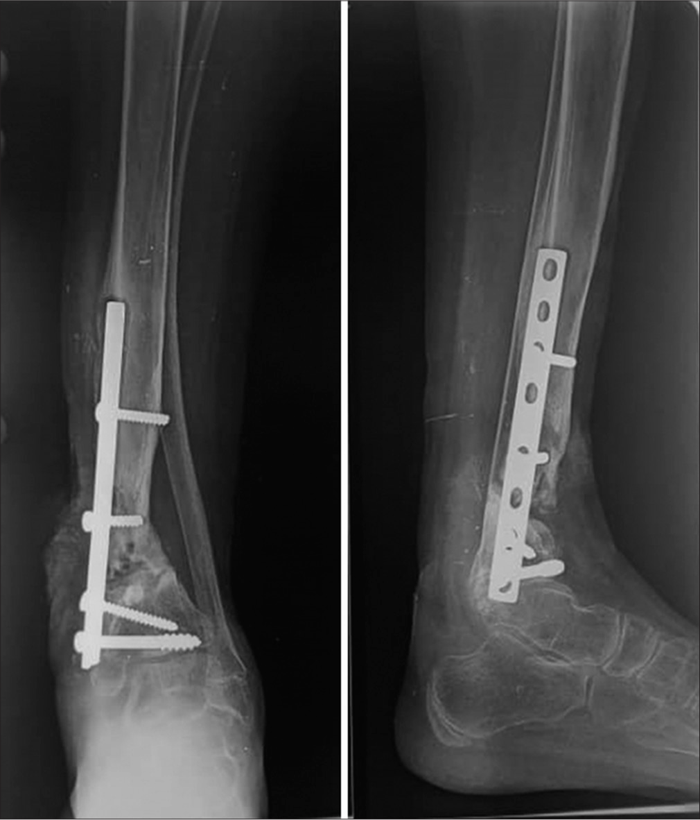
- Non-union of tibia and epiphysiodesis of distal growth plate and single growth of the fibula after plate osteosynthesis. A child of 14-years-old.
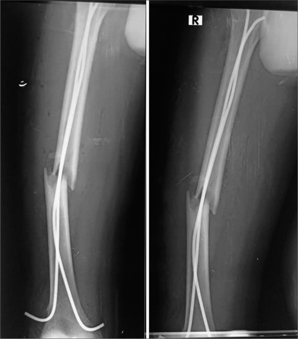
- Diaphyseal non-union of femur after elastic stable intramedullary nail. A child of 13 years old. Non-union was caused by early walking (though the nail diameter was smaller than optimal).
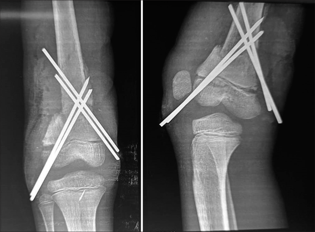
- Beginning of K-wire migration and malunion. A child of 10 years old.
Plate osteosynthesis has very limited indications in the treatment of fractures in children. According to those authors, the plates are used less and are in the process of being abandoned.[3] Using plate osteosynthesis can remove the fracture hematoma and compromise the anatomical integrity of the periosteum. It is used for certain fractures of the femur, humerus, and radius to obtain perfect anatomical reduction and, above all, to allow early mobilization. Plate osteosynthesis has been used to achieve complete consolidation of highly displaced fractures without complication.[5]
Half of all plate osteosynthesis resulted in post-operative infection [Table 4]. This finding reinforces the argument that infection is the main complication of plate osteosynthesis in children.[4] Plates should always be removed within 8–12 months postoperatively.[4,5] Osteosynthesis using ESIN has been associated with fewer complications than plate osteosynthesis. Malunion can be a complication of inadequate conservative treatment (insufficiently molded plaster with excess padding, imperfect reduction, etc.).[8] Malunion is the result of conservative treatment.[4] In this study, we found a small proportion (4.1%) of malunion [Table 4] that did not result in limb length discrepancy (LLD).
| Bone | Infection | Implant migration | Non-union | Malunion | Total |
|---|---|---|---|---|---|
| ESIN | 2 | 13 | 0 | 2 | 17 |
| Plate osteosynthesis | 11 | 1 | 8 | 1 | 21 |
| Total | 13 | 14 | 8 | 3 | 36 |
ESIN: Elastic stable intramedullary nail.
Another post-operative complication of diaphyseal fractures, which is not uncommon in the literature, is LLD. We did not find that in this study, our follow-up was not long enough to find all cases of LLD. In addition, LLD is a complication of conservative treatment. Lutz believe that LLD is exclusive to diaphyseal fractures (whatever the treatment), either by hypergrowth or malunion with or without angulation.[4] They also believe that LLD is the main sequela of femur fractures in children.[4]
Non-union is another complication that is rarely encountered in children [Table 4].[6,13,14] Like other authors, we have noted that non-union is caused by loss of bone substance or complex fractures.[6] The children in whom we found cases of non-union were all over six years of age. The locations of the non-union were all diaphyseal. Furthermore, Lascombes et al. noted that delays and defects in healing were observed exclusively in diaphyseal fractures and none of these complications were observed in metaphyseal fractures.[6]
According to the Oestern and Tscherne criteria, the outcomes of 24 fractures were excellent, 35 were good, and 15 were poor. Using the same criteria, Abiome et al. found 37 excellent and five good outcomes.[2] We assume that, unlike us, they did not get poor outcomes. This comparison could be explained by differences in the quality of the work and the assessment of the criteria. The quality of their osteosynthesis outcomes is much better than ours because they have experienced surgeons, better working materials, and good-quality orthopedic implants.
Finally, in our study, 20% of the parents were very satisfied with the care provided, 62% were satisfied, and 17% were disappointed. These subjective figures enabled us to assess the efficiency of our services.
Limitations of the study
Our study’s small sample size did not allow us to perform more convincing statistical tests. In addition, since our study was not funded, we could not conduct excellent scientific investigations.
CONCLUSION
It must always be remembered that any conservative treatment leads to faster and safer consolidation than any osteotomy. Osteosynthesis in routine pediatric traumatology is only indicated if the remodeling capacity of a callus is insufficient. Osteosynthesis in children produces good outcomes. Postoperative complications are common and often encouraged by a precarious environment. Complications are most often induced by plate osteosynthesis. The preferred surgical treatment for closed diaphyseal fractures should now be ESIN unless otherwise indicated. In all cases, regular clinical and radiological monitoring is essential for the diagnosis of early complications.
Recommendation
Further studies with a considerable sample size need to be undertaken to better define the limits between the surgical and conservative treatment of diaphyseal fractures in children.
ACKNOWLEDGMENTS
The authors would like to thank all the staff, who contributed to the management and follow-up of the patients.
AUTHORS’ CONTRIBUTIONS
HVF and DP conceived and designed the study, conducted research, provided research materials, and collected and organized data. MM and NM analyzed and interpreted data. HVF wrote the initial and final draft of the article and provided logistic support. VNN corrected the first version of the article. SRB corrected the list version of the article and supervised. All authors have critically reviewed and approved the final draft and are responsible for the manuscript’s content and similarity index.
ETHICAL APPROVAL
The Institutional Review Board has waived the ethical approval for this study.
DECLARATION OF PATIENT CONSENT
The authors certify that they have obtained all appropriate patient consent forms. In the form, the patients’ parents have given their consent verbally for their images and other clinical information to be reported in the journal. The patients’ parents understand that their names and initials will not be published, and due efforts will be made to conceal their identity, but anonymity cannot be guaranteed.
USE OF ARTIFICIAL INTELLIGENCE (AI)-ASSISTED TECHNOLOGYFOR MANUSCRIPT PREPARATION
The authors confirm that there was no use of artificial intelligence (AI)-assisted technology for assisting in the writing or editing of the manuscript and no images were manipulated using AI.
CONFLICTS OF INTEREST
There are no conflicting relationships or activities.
FINANCIAL SUPPORT AND SPONSORSHIP
This study did not receive any specific grant from funding agencies in the public, commercial, or not-for-profit sectors.
References
- Feasibility of an emergency reduction protocol for diaphyseal fractures of both forearm bones in children. Rev Chir Orthop Traumatol. 2015;101:390-3.
- [CrossRef] [PubMed] [Google Scholar]
- Surgical treatment of forearm fractures in children at Owendo University Hospital: A study of 42 cases. Health Soc Dis. 2022;23:52-4.
- [Google Scholar]
- Plate inside of bone cortex of femur removed at 21 years postoperatively: About a case. JACCR Afr. 2021;5:145-9.
- [Google Scholar]
- Submuscular bridge plating in the treatment of unstable femur fractures in children and adolescents. J Musculoskelet Surg Res. 2019;3:286-91.
- [CrossRef] [Google Scholar]
- Early complications when elastic intramedullary nailing is used for fractures in children: About 100 cases treated with pre-curved tip and stem nails. Rev Chir Orthop Trauma. 2012;98:327-34.
- [CrossRef] [PubMed] [Google Scholar]
- Thirty-five years of elastic stable intramedullary nailing (ESIN) in pediatric fractures: A method still young. E Mem Natl Acad Surg. 2015;14:109-14.
- [Google Scholar]
- Complications in operatively managed pediatric femoral shaft fractures. J Musculoskelet Surg Res. 2024;8:47-52.
- [CrossRef] [Google Scholar]
- Open-focus treatment of diaphyseal femur fractures in children by elastic and stable intramedullary pinning at Yopougon University Hospital. Rev Chir Orthop Trauma. 2015;101:385-8.
- [CrossRef] [Google Scholar]
- Evaluation of the treatment of femoral shaft fractures in children by elastic stable intramedullary nailing. Dakar Méd. 2004;49:162-6.
- [Google Scholar]
- The elastic stable intramedullary nailing (ESIN) Rev Chir Orthop Trauma. 2010;35:193-203.
- [CrossRef] [Google Scholar]
- Stable elastic intramedullary pinning of diaphyseal tibial fractures in children. J Trauma Sport. 2009;26:85-90.
- [CrossRef] [Google Scholar]
- Instability, non-union, and subsequent failure of flexible nails: Takeaways from the complication. J Musculoskelet Surg Res. 2024;8:181-3.
- [CrossRef] [Google Scholar]


