Translate this page into:
Thorn-like exostoses: A rare presentation of hereditary multiple exostoses
2 Department of Pathology, Government Medical College, Haldwani, Uttarakhand, India
Corresponding Author:
Ganesh Singh Dharmshaktu
Department of Orthopaedics, Government Medical College, Haldwani - 263 139, Uttarakhand
India
drganeshortho@gmail.com
| How to cite this article: Dharmshaktu GS, Pangtey T, Bhandari SS. Thorn-like exostoses: A rare presentation of hereditary multiple exostoses. J Musculoskelet Surg Res 2018;2:137-138 |
Dear Sir,
We read the article “Atypical presentation of hereditary multiple exostoses (HME) (osteochondromas). Thirteen years of follow-up and analysis” with great interest and it describes an uncommon behavior of exostoses.[1] Exostoses can be pedunculated or sessile or a mix of both in the settings of HME. We would like to report a rare deviant form of exostoses in a case of HME. A 23-year-old case of HME with positive family history presented to us with multiple exostoses chiefly in bilateral upper and lower extremities. The lesion in the right knee region on radiograph showed two thorn-like exostoses over the medial aspect of distal femur among other sessile lesions [Figure - 1]a. The lesions on other sites were mostly sessile and not affecting activities of daily living. He was advised periodic review and managed conservatively. One year later, he presented to us with pain over the medial aspect of the knee during movement while the full flexion was not possible due to the presence of adjacent exostoses. The radiograph at this time showed a more sharpened appearance of the previous exostoses that almost resembled thorn-like projections [Figure - 1]b. These projections might be irritating vastus medialis obliqus and surrounding region during cyclic motion. There was adjacent posterior femur exostosis that was causing flexion block, and the patient was advised a surgical excision of symptomatic lesions. The excision of the distal femoral lesion along with the thorn-like exostoses was done. The pointed lesions were sharp bony spur-like structures without presence of cartilage cap [Figure - 2]. The histopathological report of other lesions sent along with thorns complied with the diagnosis of exostoses while the thorns had no cartilage cap and were composed of cortical bony tissue. No complication or recurrence in perioperative period and follow-up was noted.
![[Figure - 1]](#fig_SaudiOrthopJ_2018_2_3_137_235506_f1.jpg)
![[Figure - 2]](#fig_SaudiOrthopJ_2018_2_3_137_235506_f2.jpg)
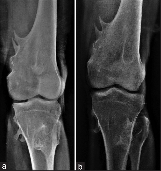 |
| Figure 1: The anteroposterior radiograph of the multiple exostoses around the knee with thorn-like lesions over the medial aspect (a), and the sharpening of the same lesions over the time (b) |
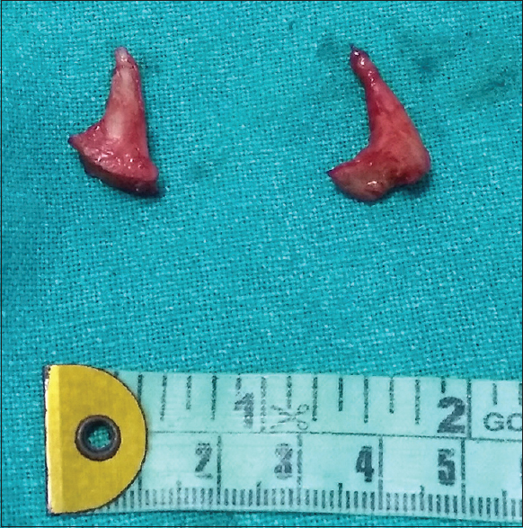 |
| Figure 2: The excised specimen of the lesion showing pointed, sharp thorn-like structures |
The presence of sharp thorn-like exostosis is an uncommon presentation with very few reports in the literature. We could find similar reports with respect to thorn-like rib exostosis causing thoracic hemorrhage.[2] Before that only four cases in children have been reported with similar lesion complicating into chest injuries.[3],[4],[5],[6] We could not find reports of similar lesion in extremities and the case highlights the importance of the evaluation of other sites for the presence of small thorn-like lesions as they may present with life-threatening complications as described in published reports of costal exostoses. Another important feature we noted was sharpening of existing lesions with time probably with the regular physiological motions. This fact should be remembered while reviewing dormant or benignly sharp lesions in the future. It is, therefore, our belief that these lesions should be addressed surgically to avoid complications arising from their mechanical effects on surrounding structures in selected cases.
Declaration of patient consent
The authors certify that they have obtained all appropriate patient consent forms. In the form the patient(s) has/have given his/her/their consent for his/her/their images and other clinical information to be reported in the journal. The patients understand that their names and initials will not be published and due efforts will be made to conceal their identity, but anonymity cannot be guaranteed.
Financial support and sponsorship
Nil.
Conflicts of interest
There are no conflicts of interest.
| 1. | Mukhtar IA, Alqasim E, Abdulkareem I, Prabhu J. Atypical presentation of hereditary multiple osteochondromas: Thirteen years of follow-up and analysis. J Musculoskelet Surg Res 2018;2:73-6. [Google Scholar] |
| 2. | Cheng PG, Chen CC, Wu SK, Hsu SM, Wang MN. Thorn-like osteochondroma presenting as hemothorax in an adult. Formos J Surg 2012;45:97-9. [Google Scholar] |
| 3. | Teijeira FJ, Baril C, Younge D. Spontaneous hemothorax in a patient with hereditary multiple exostoses. Ann Thorac Surg 1989;48:717-8. [Google Scholar] |
| 4. | Reynolds JR, Morgan E. Haemothorax caused by a solitary costal exostosis. Thora×1990;45:68-9. [Google Scholar] |
| 5. | Tomares SM, Jabra AA, Conrad CK, Beauchamp N, Phoon CK, Carroll JL. Hemothorax in a child as a result of costal exostosis. Pediatrics 1994;93:523-5. [Google Scholar] |
| 6. | Simansky DA, Paley M, Werczberger A, Bar Ziv Y, Yellin A. Exostosis of a rib causing laceration of the diaphragm: Diagnosis and management. Ann Thorac Surg 1997;63:856-7. [Google Scholar] |
Fulltext Views
5,312
PDF downloads
1,280


